header-bg

National Institute on Aging (NIA)

Quantitative imaging and pooled CRISPRi screening of single cells to understand transcription factor signaling dynamics

Project

Quantitative imaging and pooled CRISPRi screening of single cells to understand transcription factor signaling dynamics

Project Details

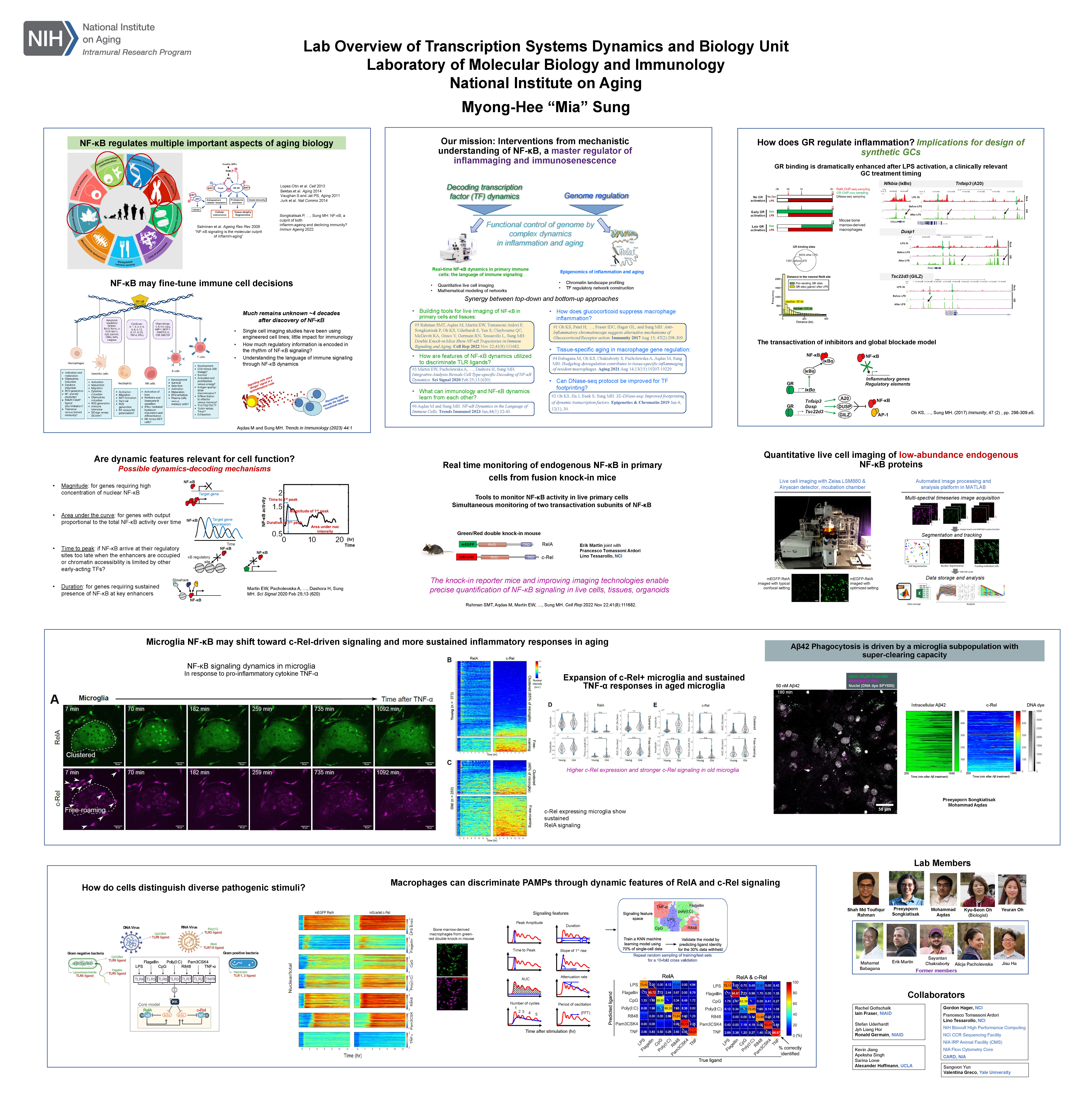
Quantitative imaging and pooled CRISPRi screening of single cells to understand transcription factor signaling dynamics. NF-κB is a master regulator of inflammation, immunity, and cell stress responses. The temporal dynamics of NF-κB signaling capture pathogen-specific information and govern the corresponding gene expression patterns. Recent studies have revealed that distinct NF-κB signaling profiles can lead to specific epigenetic modifications within potential enhancer regions of the genome, potentially establishing epigenetic memory for subsequent infections.

In this project, our goal is to investigate the impact of epigenetic perturbations on NF-κB signaling dynamics. We will utilize CRISPRi and a pool-genetic approach to systematically disrupt all potential enhancer regions around NF-κB-regulated genes. Additionally, we will conduct live-cell microscopy to quantitatively measure the resulting changes in NF-κB signaling dynamics. The insights gained from this study will illuminate the functions of the enhancer regions of NF-κB-regulated genes and will provide information on how tissue-specific NF-κB signaling is shaped by the epigenome, through the formation of epigenetic memories. The student will learn various interdisciplinary methods involving cell culture, quantitative microscopy, fluorescent reporter assays, automated single-cell analysis, molecular biology, and imaging data analysis.
Category
Institute or Center
Project Listed Date

Using long read sequencing to explore the genomic causes of the major neurodegenerative diseases

Project

Using long read sequencing to explore the genomic causes of the major neurodegenerative diseases

Project Details

Over the last 10 years, genome wide association studies (GWAS), exome and short-read genomic sequencing have enabled a revolution in our understanding of the genetic basis of neurodegenerative diseases, their progression and disease pathways. Despite this progress, our molecular understanding of the genes and loci that cause neurodegeneration remain limited, evidenced by the near absence of disease-modifying treatments for these diseases. In part this is because we have lacked the technology to fully characterise these important genomic regions. Short reads cannot fully assemble complex genomic rearrangements especially repetitive sequences, nor can they accurately and unambiguously identify or quantify different expressed isoforms. Therefore, the hypothesis underlying this PhD project is that significant inaccuracies in our knowledge of the genomic structure and transcript annotations at neurodegenerative disease loci have limited our understanding of disease pathogenesis.

To address this knowledge gap, the student will generate and analyse high quality paired long-read DNA and RNA-sequencing data to accurately investigate and annotate loci of interest in human brain samples, and in purified cell populations, across a range of neurodegenerative diseases. The student will use this new genomic and transcriptomic map to re-assess both GWAS risk SNPs at these loci and the pathogenicity of rare variants identified through WGS of patients with hereditary forms of neurodegeneration, so leveraging data generated within this project and that already available publicly. Thus, the student will help generate a core resource of annotated pathogenic loci to drive the identification of novel disease mechanisms, genetic causes and therapeutic targets in neurodegeneration.

Category
Institute or Center
University
8
Project Listed Date
UK Mentor

Characterising changes in median eminence myelination across the spectrum of body adiposity using advanced quantitative magnetic resonance imaging

Project

Characterising changes in median eminence myelination across the spectrum of body adiposity using advanced quantitative magnetic resonance imaging

Project Details

Extensive work from the Blouet lab has recently characterised the high level of myelin plasticity in the median eminence (ME), with rapid local turnover of myelin in healthy adult rodents. The ME is a region of the hypothalamus essential for various homeostatic functions, neuroendocrine output and energy balance regulation. Both weight loss, achieved through caloric restriction, and weight gain, obtained by feeding with a high fat diet, reduce ME myelin turnover, leading to local hypo- or hypermyelination, respectively. However, the contribution of changes in ME myelin plasticity and myelination to the behavioural, metabolic, or neuroendocrine adaptations engaged during energetic challenges remains unclear and how these adaptations might be impaired in aging is unknown. Investigating whether similar changes occur in humans requires novel strategies to image ME myelin in vivo in humans with high resolution and sensitivity. In this project, we propose to develop advanced magnetic resonance imaging (MRI) methodologies to perform longitudinal quantifications of ME myelination in young or aged rodents exposed to a variety of genetic or environmental perturbations modifying energy balance and adult myelin plasticity. We will also translate protocols to image and quantify ME myelin in human participants and determine the effect of age and variations across the spectrum of body mass index on ME myelin density. This project will benefit from the expertise available in Dr. Bouhrara in myelin imaging using advanced MRI methodologies to quantify ME myelination in the rodent brain in vivo and in human participants with high neuroanatomical resolution and sensitivity. These optimized protocols will be used in the Blouet lab to investigate long term changes in myelination during homeostatic and metabolic challenges. This is a unique opportunity to bridge the gap between molecular neuroscience and MR physics to address outstanding mechanistic questions regarding metabolic dysfunctions and myelination patterns. We expect that this synergetic work will form the basis for further preclinical investigations and clinical trials of targeted metabolic interventions. 

Category
Institute or Center
University
8
Project Listed Date

Predicting imaging phenotypes from multi-dimensional spectral measurements of MRI-relevant tissue properties

Project

Predicting imaging phenotypes from multi-dimensional spectral measurements of MRI-relevant tissue properties

Project Details

The past decade has seen the emergence of population-level magnetic resonance imaging (MRI) studies like the UK Biobank, which is scanning an unprecedented 100,000 individuals. This imaging has enormous potential to inform about early pathology or susceptibility to disease. However, to translate insights from population-level health data resources into the clinic, we require approaches to translating, or ‘harmonising’, between datasets acquired under very different conditions.

A newly funded collaboration between Oxford and the NIH aims to deliver a novel harmonisation approach by linking relevant tissue biology to the physics of the imaging measurement. Core to this ‘biophysical’ approach is a framework for predicting imaging phenotypes from multi-dimensional spectral measurements of MRI-relevant tissue properties.

This DPhil project will deliver the multi-spectral measurements at the heart of this prediction framework. The student will work within our collaborative team to:

  • Year 1: implement multi-spectral acquisition protocols and associated analysis pipelines for use in a large cohort;
  • Years 2-3: develop novel biophysical modelling that enables us to characterise and restrict the number of modelled tissue compartments, enabling fewer measurements for clinical scanners;
  • Year 4: demonstrate the ability to predict imaging phenotypes based on these measurements in order to harmonise measurements from multiple protocols.


This project would be jointly supervised by the neuroimaging experts in Oxford who are leading brain MRI in UK Biobank (Miller) and physics experts at NIH who have pioneered these multi-spectral measurements (Benjamini).

Institute or Center
University
7
Project Listed Date
NIH Mentor
UK Mentor

Disease pacemaker Stem Cells in Neurodegenerative Disease

Project

Disease pacemaker Stem Cells in Neurodegenerative Disease

Project Details

The presence and role of neural stem cells (NSCs) in the adult human brain is a long-debated issue in neuroscience. Recent work has demonstrated that stem-like cells exist in the embryonic, foetal, and human adult brain where they persist well into adulthood and can even contribute to neurogenesis. However, their role in neurodegenerative disease is unknown. Ongoing work in the lab has led to the hypothesis that NSCs may become dysfunctional in neurodegenerative disease resulting in senescence chronic inflammation, and thereby acting as pacemaker cells driving neuronal demise. This ambitious project aims to identify disease-associated NSCs and their phenotype in the context of human neurodegeneration using spatial biology approaches, including imaging mass cytometry, RNA scope and single nuclear RNA sequencing. Relying on post-mortem brain tissue of different stages of Alzheimer’s disease, traumatic brain injury, vascular dementia and chronic stroke, this project will study NSCs in a range of human diseases characterised by neurodegeneration and neuronal injury. Ongoing work in the lab identifies NSC-specific markers based on transcriptomics and protein profiling experiments in brains with progressive multiple sclerosis, enabling to investigate the distribution of NSCs in a wide range of diseases. Spatial transcriptomics and proteomic approaches will allow to study their phenotype and dysfunction in relation to other cell types and local pathology. This project will shed light on the role of NSCs in neurodegeneration and has the potential to identify an entirely novel mechanism of neurodegeneration in human disease.

This project will be co-supervised by Prof. A. Quaegebeur.

Institute or Center
University
8
Project Listed Date
NIH Mentor

Stem cells of the aging MS brain

Project

Stem cells of the aging MS brain

Project Details

Primary progressive multiple sclerosis (PPMS) is a chronic demyelinating disease of the central nervous system, which currently lacks restorative therapies. Transplantation of neural stem cells (NSCs) has been shown to promote healing of the injured CNS, but previous work has demonstrated that NSCs from patients with PPMS are prematurely senescent. Cellular senescence causes a pro-inflammatory cellular phenotype that impairs tissue regeneration. Senescence in PPMS NSCs was found to be associated with increased secretion of HMGB1, a pro-inflammatory alarmin found to inhibit oligodendrocyte differentiation, and also found increased within white matter lesions of PPMS autopsy tissue. This project aims to understand the role of HMGB1 in PPMS NSC senescence using techniques such as CRISPR-Cas9, RNA sequencing, and functional NSC assays. The longterm goal of this project will be to determine the cause of senescence in NSCs from patients with PPMS and if these cells are suitable for therapeutic use.

Category
Institute or Center
University
8
Project Listed Date
Back to Top