header-bg

Research Opportunities

Background Header
Image
Prospective Students

The goal of the NIH Oxford-Cambridge (OxCam) Scholars Program is to create, foster, and advance unique and collaborative research opportunities between NIH laboratories and laboratories at the University of Oxford or the University of Cambridge. Each OxCam Scholar develops a collaborative research project that will constitute his/her doctoral training. Each Scholar also select two mentors – one at the NIH and one in the UK – who work together to guide the Scholar throughout the research endeavor.

Students may select from two categories of projects: Self-designed or Prearranged. OxCam Scholars may create a self-designed project, which enables students to develop a collaborative project tailored to his/her specific scientific interests by selecting one NIH mentor and one UK mentor with expertise in the desired research area(s). Alternatively, students may select a prearranged project provided by NIH and/or UK Investigator(s) willing to mentor an OxCam Scholar in their lab.

Self-designed Projects 
Students may create a novel (or de novo) project based on their unique research interests. Students have the freedom to contact any PI at NIH or at Oxford or Cambridge to build a collaboration from scratch. The NIH Intramural Research Program (IRP) represents a community of approximately 1,200 tenured and tenure-track investigators providing a wealth of opportunity to explore a wide variety of research interests. Students may visit https://irp.nih.gov to identify NIH PIs performing research in the area of interest. For additional tips on choosing a mentor, please visit our Training Plan.

Prearranged Projects
Investigators at NIH or at Oxford or Cambridge have voluntarily offered collaborative project ideas for NIH OxCam Scholars. These projects are provided below and categorized by research area, NIH Institute/Center, and University. In some cases, a full collaboration with two mentors is already in place. In other instances, only one PI is identified, which allows the student to select a second mentor to complete the collaboration. Please note that prearranged project offerings are continuously updated throughout the year and are subject to change.

20 Search Results

CAPTCHA
644
Category:
Cancer Biology
Project:

The link between NRF2 and BACH1 in redox pathways and radiation responses

Project Listed Date:
Institute or Center:
N/A
NIH Mentor:
N/A
UK Mentor:

Prof. Ejung Moon

University:
Oxford
Project Details:

Radiation (RT) is effective in treating many types of cancers by inducing oxidative stress through the generation of reactive oxygen species (ROS) that result in DNA damage. However, both tumour intrinsic mechanisms for suppressing ROS as well as the hypoxic microenvironment reduce the efficacy of RT. While significant efforts are being pursued to enhance radiation efficacy, our lab will focus on RT-induced ROS that kill cells by inducing ferroptosis. Ferroptosis has recently been described as a non-apoptotic form of cell death dependent on iron and lipid peroxides that contributes to radiation-induced cell death. Studies suggest that the ferroptosis pathway is independent of DSBs and that enhancing ROS induced lipid peroxidation can promote cell killing as well as overcome the problem of tumour radioresistance mediated by intrinsic radical scavengers. NRF2 is a transcription factor playing a major role in protecting cells from oxidative damage. When bound to small MAF proteins, NRF2 transcriptionally activate its target genes through binding to the antioxidative response element (ARE) on their promoter regions. BACH1 is a transcriptional repressor of these antioxidative genes through the competition with NRF2 for ARE and small MAF bindings. In the recent study suggests that NRF2 promotes BACH1-mediated lung cancer metastasis through BACH1 stabilisation. Therefore, there seems to be a tight regulation of NRF2 and BACH1 in promoting cancer through competition or cooperation. In our proposed study we will determine how NRF2 and BACH1 play together in radiation responses while focusing on redox pathways, ferroptosis, and DNA damage.

640
Category:
Cancer Biology
Project:

Investigating Novel Radiation-sensitising Drugs

Project Listed Date:
Institute or Center:
N/A
NIH Mentor:
N/A
UK Mentor:

Prof. Geoff Higgins

University:
Oxford
Project Details:

Radiation therapy is a common treatment modality for cancer patients that eliminates malignant cells through the delivery of high-energy photons. Despite advancements in radiation therapy technologies, factors such as the presence of tumour hypoxia or cell-intrinsic mechanisms of radioresistance limit the effectiveness of this treatment modality. This project aims to investigate the potential of novel drugs to enhance tumour radiosensitivity without causing toxicity to normal tissues. The research plan includes conducting in vitro and in vivo experiments using a broad panel of cancer cells to evaluate the radiosensitising effects of novel compounds which are currently being investigated in Geoff Higgins’ lab (Department of Oncology, University of Oxford). The radiosensiting capacity of these drugs and their mechanisms of action will be determined using a broad range of cell & molecular biology techniques, like colony formation assays, tumour growth delay assays, the analysis of DNA damage repair pathways by fluorescence microscopy and reporter assays, cell cycle determination by flow cytometry, gene silencing, cytogenetics assays, or protein biochemistry, amongst other techniques.

632
Category:
Cancer Biology
Project:

Systems immunology approaches to dissect the role of tertiary lymphoid structures in cancer

Project Listed Date:
Institute or Center:
N/A
NIH Mentor:
N/A
University:
Oxford
Project Details:

The formation of high-quality germinal centres (GCs) is paramount to developing antibody responses central to resolving disease. How these antibodies are generated in such an efficient and well-regulated manner relies on a controlled and compartmentalised immune-regulatory environment to prevent the production of self-reactive autoantibodies. Reduced GC function has been widely reported in infection, autoimmune diseases, and ageing. Advancing our understanding of the cellular processes curtailing the host immune-regulatory environment modulating GCs could have a clinical impact.

Over the last couple of years, evidence has emerged revealing the presence of T-cell-B-cell-rich tertiary lymphoid structures (TLS) close to tumour cells have been associated with overall survival and better response to immunotherapy in cancer, suggesting an immune benefit. Yet, their interindividual variation in cellular composition, spatial organisation, and the immune mechanisms regulating humoral responses remain unclear. 

With more than ten years of expertise in the HIV field with a focus on the biological processes underpinning the regulation of humoral responses, the Functional Immunology lab led by Dr Pedroza-Pacheco aims to translate their established methodologies to systematically quantify the functional relationship between tumour-intrinsic molecular processes, and the formation, cellular composition, and spatial distribution of CD4-B-cell-rich TLS within the tumour microenvironment. Understanding how CD4 Tfh and B cells contribute to anti-tumour responses provides an exciting opportunity for their translation into precision immunotherapies, non-invasive biomarkers, and cancer vaccines. 

588
Category:
Cancer Biology
Project:

Understanding cancer clonal dynamics towards novel therapeutic approaches

Project Listed Date:
Institute or Center:
National Cancer Institute (NCI)
NIH Mentor:

Dr. Sam Mbulaiteye

University:
Cambridge
Project Details:

Burkitt lymphoma (BL) is an aggressive cancer of germinal centre B cells that largely affects children globally. In sub-Saharan Africa, Burkitt lymphoma is an endemic disease associated with Epstein-Barr Vius (EBV) and Plasmodium falciparum infection. Unfortunately, children in sub-Saharan Africa have a far worse outcome with about 40% of children surviving compared to greater than 90% elsewhere, particularly in high income countries in Europe and North America. This is due to low access to reliable pathology diagnosis, limited access to specialized oncology centres, where the effective cytotoxic treatments and necessary life support can be given to patients during care. However, there might be biological factors that contribute as well to differences in outcome> For example, Burkitt lymphoma in sub-Saharan Africa is associated with EBV and Plasmodium falciparum infection, which may mediate a different tumour landscape (predominated by action of mutator enzyme adenosine-induced cytosine deaminase), whereas elsewhere these factors are lacking and the tumour landscape is influenced by accumulation of mutations in genes influencing apoptosis. In Cambridge, Prof. Turner has developed in vivo models of both sporadic and endemic Burkitt lymphoma that facilitate comparative research into disease mechanisms. In this project, these will be employed to understand the clonal heterogeneity of these malignancies using a combination of in vivo CRISPR screens and lineage tracing. Data will be validated using a large resource of primary patient specimens available within the EMBLEM study coordinated by the National Cancer Institute. Ultimately, data will be analysed with a view to developing biomarkers of disease prognosis as well as novel therapeutic approaches. In both cases the resource settings of sub-Saharan Africa will be considered towards sustainable and achievable approaches. The student will have the opportunity to travel to Uganda during the course of their studies.

459
Category:
Cancer Biology
Project:

Understanding the XPO7:SLK complex to formulate synergistic combination therapies

Project Listed Date:
Institute or Center:
National Cancer Institute (NCI)
UK Mentor:

Prof. Jon Elkins

University:
Oxford
Project Details:

Most translational efforts for uncommon cancers stem from oncogenic mutations identified by sequencing. In contrast to genomic analyses, the tumor proteome has the potential to better approximate phenotype-inducing alterations, particularly in the absence of targetable driver mutations. However, tumor analyses using mass spectrometry can be challenging. Exosomes (small extracellular membrane-enclosed vesicles) may circumvent these issues and serve as a valuable, prioritized, “window” into the tumor cell proteome. Applying this reasoning to bile duct cancers (cholangiocarcinoma, CCA), we performed mass spectrometry on exosomes extracted from patient bile, revealing a 17-fold enhancement of the nuclear export protein XPO7. Immunohistochemistry analysis of XPO7 expression in 318 CCA patients unexpectedly demonstrated intense cytoplasmic staining. Within the cytosol we demonstrate that XPO7 exists in a molecular complex with the serine/threonine kinase SLK. shRNA-mediated knockdown of either XPO7 or SLK in CCA lines abrogated tumor organoid formation and reduced orthotopic tumor growth. To translate the target to patients, we identified tivozanib as a potent SLK inhibitor. Tivozanib treatment reduced tumor organoid formation in vitro and induced tumor regression in vivo in patient derived xenografts (n=2). Together, these findings reveal a novel cytosolic XPO7:SLK signaling axis that is targetable in CCA patients and we have already documented early responses with our accruing Phase I/II trial (NCT 04645160). It is however clear that single agents will not result in cures for patients with solid tumors, and a better understanding of the XPO7:SLK complex (including downstream oncogenic signaling axes) will be required to formulate and implement synergistic combination therapies.

434
Category:
Cancer Biology
Project:

Exploring the relationship of transient blood-brain barrier disruption to inhibition of malignant glioma progression

Project Listed Date:
Institute or Center:
National Institute of Neurological Disorders and Stroke (NINDS)
NIH Mentor:

Dr. Sadhana Jackson

UK Mentor:
N/A
University:
N/A
Project Details:

One of the major obstacles to effectively treating central nervous system (CNS) tumors is the integrity of the blood-brain barrier (BBB). The BBB prevents systemic drug delivery from reaching the brain and brain tumor tissue. While previous studies have mainly focused on circumventing the BBB, very few agents or mechanisms have been explored that modulate the tumor microenvironment to enhance effective therapies for malignant brain tumors. Our studies focus on understanding the heterogeneity of BBB permeability amongst malignant tumor cells and the role of the supportive BBB in tumor growth. Our collaborative laboratory and clinical investigations center around BBB biology, cancer biology, pharmacokinetics and pharmacodynamics related to optimal CNS drug delivery.

Using a clinical/translational approach, we aim to:

1) Evaluate the efficacy of targeted tumor and BBB directed therapy

2) Define the mechanisms that drive differences in neuropharmacokinetics of agents to the CNS

3) Identify exquisite parameters via neuro-imaging of CNS permeability amongst malignant brain tumors.

Our overall goal is to enhance our understanding of the heterogeneity of blood-brain barrier permeability among tumor cells and develop mechanism-based therapeutic interventions to treat affected brain tumor patients at the NIH Clinical Center. We use a combination of cell biology, molecular biology, imaging, pharmacokinetics and animal tumor models.

353
Category:
Cancer Biology
Project:

Metabolic regulation of gene expression in the context of cancer

Project Listed Date:
Institute or Center:
National Cancer Institute (NCI)
NIH Mentor:

Dr. Len Neckers

University:
Cambridge
Project Details:

Emerging evidence suggests an exciting link between metabolism, chromatin and transcription. Metabolism can regulate post-translational modifications of histones which in turn regulate transcription of target genes. Highly proliferative cancer cells re-wire their metabolism to fuel growth, and in turn modify histones to alter gene expression. Identifying mechanisms by which cancer cells re-wire their metabolism and gene expression will identify key vulnerabilities to target using small molecule therapeutics.

Our recent work at NIH has demonstrated links between histone lactylation, gene expression and cancer metabolism (histone lactylation depends on elevated cellular lactate, the end product of glycolysis – a preferred metabolic pathway in cancer). Work in Cambridge has further linked the molecular chaperone HSP90 with gene expression and metabolism in the context of cancer. Harnessing the complementary strengths in the two labs at NIH and Cambridge, the collaborative work will delineate molecular pathways linking small-molecule therapeutics targeting the chaperone HSP90 with cancer metabolism and with specific small-molecule inhibitors of glycolysis. The data we obtain delineating the metabolic dependence of gene expression in cancer will uncover novel and exciting treatment strategies to treat cancers’ metabolic vulnerabilities.

228
Category:
Cancer Biology
Project:

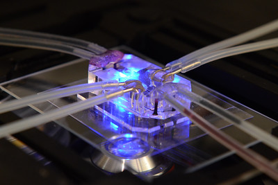
Establish and implement a glioblastoma-on-a-chip model to study the effect of microenvironments on the tumor progression

Project Listed Date:
Institute or Center:
N/A
NIH Mentor:
N/A
University:
Cambridge
Project Details:
N/A
197
Category:
Cancer Biology
Project:

Crosstalk between the tumour suppressor p53 and inflammation pathways

Project Listed Date:
Institute or Center:
N/A
NIH Mentor:
N/A
UK Mentor:

Prof. Xin Liu

University:
Oxford
Project Details:
N/A
185
Category:
Cancer Biology
Project:

What types of physical activity are associated with a lower incidence of cancer?

Project Listed Date:
Institute or Center:
National Cancer Institute (NCI)
NIH Mentor:

Dr. Charles Matthews

UK Mentor:

Prof. Aiden Doherty

University:
Oxford
Project Details:

At the National Cancer Institute, we have demonstrated that higher levels of moderate to vigorous intensity physical activity are associated with a lower risk of cancer, including cancer in the breast, colon, endometrium, bladder, kidney, and stomach1. However, due to a reliance on self-reported measures of physical activity, a number of key questions remain unanswered on what overall volume of physical activity, and what types of physical activity, are associated with lower cancer risk. In addition, previous studies are observational by nature and are therefore unable to determine causality due to unmeasured or residual confounding.

At Oxford, our group has shown that wearable sensors such as wrist-worn accelerometers can be used to noninvasively measure physical activity status in large-scale biomedical studies. For example, we have measured physical activity status in 103,712 UK Biobank participants who agreed to wear a wrist-worn accelerometer for seven days2. These measurements are now actively used by health researchers worldwide to demonstrate that simple measures of overall activity are cross-sectionally associated with cancer outcomes3. However, no large study of device measured physical activity has yet taken place to assess associations with incident cancer outcomes with sufficient longitudinal follow-up. Furthermore, activity trackers often capture ~180 million data points/participant/week and therefore have the potential to identify other powerful behavioural signals to detect future cancer risk.

Machine learning methods can help maximise the utility of data from wearable sensors. These methods attempt to automatically detect patterns in data and then use those uncovered patterns to predict future data. Our group has demonstrated the utility of supervised machine learning to identify sleep and functional physical activity behaviours from raw accelerometer data4. However, there is a broad concern around the lack of reproducibility of machine learning models in health data science5. It is therefore important to carefully consider how to promote robust machine learning findings and reject irreproducible ones, to ensure credibility and trustworthiness.

This DPhil project therefore proposes to use the world’s largest available datasets to investigate what types of physical activity are associated with a lower incidence of cancer. Working with colleagues at the University of Oxford and the National Cancer Institute, you will have the opportunity to address the following important questions:

1. What behavioural measurements of physical activity status can be reliably ascertained from accelerometer datasets?
You will have the opportunity to develop reproducible machine learning skills to develop methods to identify physical activity behaviours from raw accelerometer datasets. Specifically, you will develop semi-supervised machine learning methods which seek to combine supervised methods (good quality labels, small datasets) with unsupervised methods (no labels but large datasets which are less prone to sampling bias). This will involve use of the largest available accelerometer datasets with reference measurements for physical activity behaviours in free-living environments (using wearable cameras)6.

2. What physical activity behaviours are associated with incident cancer events?
Here, you will have the opportunity to develop new skills in epidemiological data analysis. You will have the opportunity to use the UK Biobank dataset which has collected wrist worn accelerometer data from 103,712 participants2. This dataset includes information on participants’ first hospital admission or death from cancer, identified from linkages to the national death index, Hospital Episode Statistics, and cancer registries.

3. Are physical activity behaviours potentially causally associated with cancer?
You will have the opportunity to develop genetic epidemiology skills by implementing two-sample Mendelian Randomization7 to assess potential causal effects of accelerometer measured physical activity and cancer. For cancer outcomes, summary genetic association data will be obtained from existing collaborators from International cancer consortia.

Candidates should have a BSc, or ideally MSc, in a discipline with a substantive epidemiological, computational, or quantitative component. We very much welcome prospective candidates to directly contact us to further develop this proposal.

166
Category:
Cancer Biology
Project:

Identifying Regulators of Cancer Stem Cells in Pancreatic Cancer

Project Listed Date:
Institute or Center:
National Cancer Institute (NCI)
NIH Mentor:

Dr. Udo Rudloff

UK Mentor:

Prof. Siim Pauklin

University:
Oxford
Project Details:

Pancreatic ductal adenocarcinoma (PDAC) is one of the most lethal malignancies in human due to its late detection, highly metastatic characteristics, and poor responsiveness to current therapeutics. Pancreatic tumorigenesis involves a dedifferentiation process of cellular identity and the acquisition of a stem cell-like state of a subpopulation of cells known as cancer stem cells (CSCs). These cells are exceptionally important due to their higher therapeutic resistance and phenotypic plasticity that allows CSCs to metastasize and give rise to tumours. Currently, it remains largely unclear, which molecular markers and protein machineries control the stem cell-like identity of pancreatic CSCs. This knowledge would be valuable for earlier cancer detection and for developing more efficient pancreatic cancer therapeutics in the future.


The research objective of the project is to identify and characterize novel transcriptional regulators which govern gene expression of pancreatic cancer cells, particularly stem cell-like characteristics CSCs. The project will apply a broad range of cutting-edge research techniques such as 2D and 3D human cell culture systems, co-cultures of different cell types, next-generation single cell sequencing (scRNA-seq, scATAC-seq) of tumoural subpopulations in genetically engineered murine models (GEMMs) of pancreas cancer, functional studies (CRISPR/Cas9-mediated gene editing, tumour sphere assays), mechanistic studies (confocal microscopy, flow cytometry, cell sorting, CyTOF, western blotting), patient samples and mouse in vivo studies.


Collectively, this project will provide key insights to the signalling pathways and molecular mechanisms essential for the formation and maintenance of pancreatic CSCs, helping to better understand the tumorigenic process, and to uncover novel ways for diagnosing and treating this lethal cancer.

165
Category:
Cancer Biology
Project:

Understanding combination cytotoxic chemotherapy in Acute Myeloid Leukaemia

Project Listed Date:
Institute or Center:
National Heart, Lung, and Blood Institute (NHLBI)
NIH Mentor:

Dr. Chris Hourigan

UK Mentor:

Prof. Paresh Vyas

University:
Oxford
Project Details:

Acute Myeloid Leukaemia (AML) is the most common, aggressive human leukemia. Within the whole group of AML patients there is a subset of patients, typically younger (less than 65 years of age) who receive intensive conventional combination cytotoxic chemotherapy (anthracyclines and nucleoside analogues), who have a higher cure rate (~65%). Despite these cytotoxic drugs being in routine clinical use since the 1970’s, the field surprisingly still does not understand why these patients are cured. Conventional wisdom is that these patients are cured, because intensive combination cytotoxic chemotherapy kills all AML cells. However, this has never been rigorously proven and alternative hypotheses have not been tested.

This proposal will test if in patients who are cured, compared to those who are not, if eradication of all AML cells, could result from:
1. Increased killing of AML from cytotoxic chemotherapy.
2. An autologous innate and, or, acquired immune anti-AML cell response.
3. A combination of (1) and (2).

Specific Aims:

Using patient samples from cured patients and patients who relapse we will:
1. Contrast amount of AML cells left after treatment (measurable residual disease, MRD), in bone marrow (BM) samples.
2. If residual disease is detected in samples, characterise the single cell (sc) clonal architecture, epigenome and transcriptome and determine the leukemic stem cell content of the residual AML.
3. Perform an unbiased sc transcriptomic analysis of innate and acquired immune cells in BM, and peripheral blood (PB).
4. Test functional differences in comparable immune cells.

162
Category:
Cancer Biology
Project:

Identifying sub-populations of cells critical for cancer disease progression

Project Listed Date:
Institute or Center:
National Cancer Institute (NCI)
Project Details:

Tissue multiplexing is a new imaging method that allows to visualise a large number of protein targets in tissues. This exciting new technology allows for new approaches to phenotyping cells and to decode more complex patters of communication between different tissue compartments. The goal of this project is to develop the required image analysis and inference methods using advanced machine learning and AI in 2D and 3D. As a result, you will be advancing our understanding of the tumour environment and find novel ways of identifying sub-populations of cells that play a critical role in disease progression.


You will be working side by side with world leading cancer researchers at NIH NCI and the University of Oxford. At both sites you will have access to unique patient cohorts. Together with David Wink and Stephen Lockett (both NCI) you will be working on aspects if breast cancer. In Oxford, Richard Bryant and Ian Mills will lead on work in prostate cancer, which is the commonest non-cutaneous cancer in men, and often progresses to incurable metastatic disease. Your work will also be supported by expert pathologists and you will be working towards improving current practice in cellular pathology.


The broader group has already established a very active collaboration and you will be expected to work in both locations. In Oxford, you will be embedded in the Quantitative Biomedical Image Analysis group led by Prof. Rittscher. Part of your role will be to accelerate the exchange of technology and software between the two locations. This project provides a unique opportunity to study mechanisms that are common to different cancer types.

160
Category:
Cancer Biology
Project:

Exploring mechanisms underlying heterogeneity of response in personalized cancer immunotherapy by using machine-learning techniques

Project Listed Date:
Institute or Center:
N/A
NIH Mentor:
N/A
UK Mentor:

Dr. Hashem Koohy

University:
Oxford
Project Details:

T cell recognition of a cognate peptide-MHC (pMHC) complex presented by infected/malignant antigen-presenting cells are of utmost importance for mediating a robust and long-term immune response. The recognition is mediated by specific molecular interactions between heterodimeric T Cell Receptors (TCRs) and pMHC ligands and instructs the nature of ensuing adaptive immune response. A better understanding of TCR:pMHC interaction would allow further harnessing of the adaptive T cell immunity and may lead to the development of  vaccines and therapeutics  both in the context of personalized cancer immunotherapies and infectious diseases such as COVID19. The research interests in the Koohy group are focused on the development of machine-learning and Bayesian statistical models to help us better understand two key components of this interaction: A) architecture of the immune repertoire and its dynamics upon exposure to antigens, B) processing and presentation of antigens by MHC molecules to their cognate T cells.


Cancer is usually characterized by accumulation of genetic alterations. Tumour-specific somatic mutations may generate small mutated proteins known as neoantigens that are presented on the surface of cancer cell as ‘cancerous flags’ in association with class I and II HLA molecules.  Neoantigens can be recognized by autologous T cells as foreign and therefore are considered as targets for improved cancer vaccines and adaptive T cell therapies. Almost similar mechanisms are applied to infectious diseases with the difference that the immunogenic epitopes on the surface of infected cells originate from invasive pathogens. Prediction of both immunogenic viral epitopes and cancer neoantigens has been at the centre of extensive research around the globe over the past couple of decades but remains unsolved.   We have been developing various statistical models such as Bayesian Hidden Markov Models to predict immunogenic epitopes that can be used as targets for vaccines for personalized cancer immunotherapy as well as infectious diseases such as COVID1,2.


Over the past decade we have witnessed unprecedented achievements on various cancer immunotherapies in which patients’ own immune system is modulated to find and kill cancer cells. This is evident by the 2018 Nobel Prize for development of Immune Checkpoint Blocked ICB that has greatly improved patients care. However, not all patients respond the same way, besides, some patients develop immune related adverse events such as checkpoint colitis. 
Multiple factors affect immune response to treatment including mutation burden rate, cytotoxic T cell infiltration, antigen processing and presentation defects, mutation-driven clonal signature and the composition of intestinal microbiota. Owing to advances in high throughput sequencing technologies, in particular recent single cell advancements, these features can now be measured from patients’ samples at single cell level at multiple time points including before, during and after the treatment.  We take readouts of these experiments in the form of high throughput sequencing data including genomics, transcriptomics, T cell receptor repertoire, and epigenomics data to train  statistical and machine learning models to study the mechanisms underlying heterogeneity of the response.

132
Category:
Cancer Biology
Project:

Understanding the mechanisms of tumorigenesis in individuals with predisposition to neuroendocrine tumor syndromes

Project Listed Date:
Institute or Center:
National Institute of Child Health and Human Development (NICHD)
NIH Mentor:

Dr. Karel Pacak

UK Mentor:
N/A
University:
N/A
Project Details:

Undertake genomic and epigenomic studies into the mechanisms of tumorigenesis in individuals with inherited predisposition to neuroendocrine tumor syndromes, especially pheochromocytoma/paraganglioma associated with mutations in the Krebs cycle. Such discoveries can lead to understanding of developmental and other mechanisms in these tumors related to the same syndrome but behaving in a different way and occurring in different tissue of origin. Such data can be paramount to study novel therapeutic approaches for these tumors based on the discovery on novel tumor-specific targets as well as biomarkers.

Back to Top