header-bg

Research Opportunities

Background Header
Image
Prospective Students

The goal of the NIH Oxford-Cambridge (OxCam) Scholars Program is to create, foster, and advance unique and collaborative research opportunities between NIH laboratories and laboratories at the University of Oxford or the University of Cambridge. Each OxCam Scholar develops a collaborative research project that will constitute his/her doctoral training. Each Scholar also select two mentors – one at the NIH and one in the UK – who work together to guide the Scholar throughout the research endeavor.

Students may select from two categories of projects: Self-designed or Prearranged. OxCam Scholars may create a self-designed project, which enables students to develop a collaborative project tailored to his/her specific scientific interests by selecting one NIH mentor and one UK mentor with expertise in the desired research area(s). Alternatively, students may select a prearranged project provided by NIH and/or UK Investigator(s) willing to mentor an OxCam Scholar in their lab.

Self-designed Projects 
Students may create a novel (or de novo) project based on their unique research interests. Students have the freedom to contact any PI at NIH or at Oxford or Cambridge to build a collaboration from scratch. The NIH Intramural Research Program (IRP) represents a community of approximately 1,200 tenured and tenure-track investigators providing a wealth of opportunity to explore a wide variety of research interests. Students may visit https://irp.nih.gov to identify NIH PIs performing research in the area of interest. For additional tips on choosing a mentor, please visit our Training Plan.

Prearranged Projects
Investigators at NIH or at Oxford or Cambridge have voluntarily offered collaborative project ideas for NIH OxCam Scholars. These projects are provided below and categorized by research area, NIH Institute/Center, and University. In some cases, a full collaboration with two mentors is already in place. In other instances, only one PI is identified, which allows the student to select a second mentor to complete the collaboration. Please note that prearranged project offerings are continuously updated throughout the year and are subject to change.

13 Search Results

CAPTCHA
629
Category:
Biomedical Engineering & Biophysics
Project:

The nanoscale regulation of cell surface mechanics

Project Listed Date:
Institute or Center:
N/A
NIH Mentor:
N/A
UK Mentor:

Prof. Ewa Paluch

University:
Cambridge
Project Details:

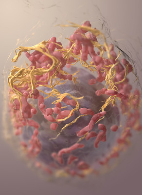
The Paluch lab investigates how cells control their shape and the underlying cellular mechanical properties. The project will focus on the actomyosin cortex, a thin cytoskeletal network that supports the plasma membrane. Myosin-generated contractility at the cell cortex controls cell surface mechanics and drives cellular deformations. Recently, through super-resolution microscopy approaches, we have shown that in interphase cells, myosin minifilaments are positioned at the cytoplasmic side of the actomyosin cortex. Upon mitotic entry, myosin minifilaments penetrate the actin cortex as cortical tension drastically increases. How this increase, crucial for the success of cell division, is controlled is not understood. We hypothesize that the cortex is structurally poised for rapid tension changes, and that tuning actin network nanoscale architecture can lead to abrupt changes in the overlap of actin and myosin at the cortex. Myosin entry into the cortex upon mitosis entry would thus be akin to a phase transition in cortex organisation.

To address this hypothesis, we will explore the 3D nanoscale architecture of myosin minifilaments at the cortex. Using Structured Illumination Microscopy, we aim to gain a single molecule understanding of the dynamic behaviour of myosin minifilaments at the onset of mitosis. We will then use Electron Microscopy to interrogate the ultrastructure of the actomyosin cortex, which together with our light microscopy data, will uncover how nanoscale processes control global cell mechanics.  

Key reference: 
Truong Quang BA, Peters R, Cassani DAD, Chugh P, Clark AG, Agnew M, Charras G, Paluch EK. Extent of myosin penetration into the actin cortex regulates cell surface mechanics. (2021) Nat Comm. 12:6511.

467
Category:
Biomedical Engineering & Biophysics
Project:

Functionalised biopolymers for regenerative and therapeutic soft robotics

Project Listed Date:
Institute or Center:
N/A
NIH Mentor:
N/A
UK Mentor:

Prof. Malavika Nair

University:
Oxford
Project Details:

In the last decade alone, degenerative diseases have featured heavily in the ten leading causes of death. Degenerative diseases and injuries have not only seen a marked increase in mortalities but are also the major contributor to the rising disability in our aging populations. As a result, there has been significant interest in developing improved implantable medical devices, which aim to replace, support, and restore the function and mobility lost by diseased tissues.

The extra cellular matrix (ECM) of tissues is an excellent base material for therapeutic and regenerative biomedical devices, since they can mimic the biological, chemical and physical environment experienced by cells in healthy tissue. However, the biomedical devices currently fabricated from the ECM have limited tunability or dynamic control once implanted within the body. The aim of this project is to develop soft robotic biomedical devices from biological polymers. Soft robots are flexible, have a high specific strength and high response rates which make them ideal for applications requiring sensitive motions. Biopolymers are an attractive material choice for biomedical soft robots: they are abundant, biodegradable, and can offer excellent biomimicry if derived from the ECM. However, these polymers typically display limited stimulus-driven shape change on their own.

As part of the project, the student will optimise the electroactivity of tissue-derived biopolymers and eventually develop a proof-of-concept therapeutic device for an application of their interest. Possible applications include (but are not limited to) drug delivery, neural and cardiac stimulators, sensors for cell attachment and proliferation. The project will involve the fabrication and structural characterisation at the nano- and micro-scale, assessment of electrical activity and opportunities for in-vitro/in-vivo testing.

446
Category:
Biomedical Engineering & Biophysics
Project:

Development of a high throughput 3D-microfluidics-based blood-brain barrier assay system as an in vitro model for ultrasound-mediated delivery of drugs into the brain

Project Listed Date:
Institute or Center:
N/A
NIH Mentor:

Dr. Marc Ferrer 

University:
Oxford
Project Details:

More than 90% of the drugs that enter clinical trials fail because of lack of efficacy or unexpected toxicity. This high failure rate has been partly attributed to the use of in vitro cellular assays and animal models that do not reproduce human physiology and pathology during preclinical drug development. This gap in clinical predictability in drug development is especially severe for CNS disorders, in which many therapeutics must cross the blood-brain barrier (BBB) to be effective. There has been a steady development and evaluation of drug delivery mechanisms that bypass the BBB to reach the target site in the CNS, and ultrasound (US)-mediated therapeutics extravasation has been one of such tools. However, while there have been several in vivo animal and clinical studies done on the focused US-guided BBB disruption, there have not yet been an established in vitro model in which US has been utilized to understand the BBB tight junction disruption and penetration. This project seeks to develop a microfluidics-based BBB model in order to: (1) investigate brain endothelial cell behaviour in microenvironment when stimulated by US and microbubbles; (2) establish whether US provides a means to enhance drug delivery through the BBB using the in vitro microfluidics-based assay platform; (3) investigate the stimulation of any inflammatory responses arising from ultrasound/microbubble exposure (in collaboration with NCI, Frank Lab). The work will contribute to greater understanding of interactions between US-mediated microbubble disruption of the BBB, especially benefitting the physicians working to utilize the technique in clinical settings and the researchers developing microbubbles to establish and optimize BBB drug delivery during the drug R&D pipeline.

437
Category:
Biomedical Engineering & Biophysics
Project:

Predicting imaging phenotypes from multi-dimensional spectral measurements of MRI-relevant tissue properties

Project Listed Date:
Institute or Center:
National Institute on Aging (NIA)
NIH Mentor:

Dr. Dan Benjamini

UK Mentor:

Prof. Karla Miller

University:
Oxford
Project Details:

The past decade has seen the emergence of population-level magnetic resonance imaging (MRI) studies like the UK Biobank, which is scanning an unprecedented 100,000 individuals. This imaging has enormous potential to inform about early pathology or susceptibility to disease. However, to translate insights from population-level health data resources into the clinic, we require approaches to translating, or ‘harmonising’, between datasets acquired under very different conditions.

A newly funded collaboration between Oxford and the NIH aims to deliver a novel harmonisation approach by linking relevant tissue biology to the physics of the imaging measurement. Core to this ‘biophysical’ approach is a framework for predicting imaging phenotypes from multi-dimensional spectral measurements of MRI-relevant tissue properties.

This DPhil project will deliver the multi-spectral measurements at the heart of this prediction framework. The student will work within our collaborative team to:

  • Year 1: implement multi-spectral acquisition protocols and associated analysis pipelines for use in a large cohort;
  • Years 2-3: develop novel biophysical modelling that enables us to characterise and restrict the number of modelled tissue compartments, enabling fewer measurements for clinical scanners;
  • Year 4: demonstrate the ability to predict imaging phenotypes based on these measurements in order to harmonise measurements from multiple protocols.


This project would be jointly supervised by the neuroimaging experts in Oxford who are leading brain MRI in UK Biobank (Miller) and physics experts at NIH who have pioneered these multi-spectral measurements (Benjamini).

436
Category:
Biomedical Engineering & Biophysics
Project:

Novel microfluidics-based technology for vaccine encapsulation

Project Listed Date:
Institute or Center:
N/A
NIH Mentor:
N/A
UK Mentor:

Prof. Anita Milicic

University:
Oxford
Project Details:

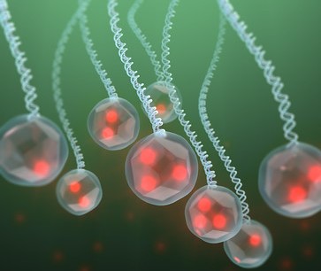
Most of the current vaccines require two or three doses for optimal efficacy. Missed booster doses contribute to incomplete immunisation coverage globally, resulting in an increased disease burden and delaying pathogen eradication. Achieving full immunisation with a single injection containing both the priming and a booster dose has been a long-standing goal in vaccinology; here the booster dose is encapsulated in polymer microparticles for a delayed release within the body after a pre-defined time period post-injection.    

We have developed a novel microfluidics-based technology for vaccine encapsulation that allows burst-release of the booster vaccine after a range of time intervals and can provide the booster immunisation in vivo. However, recent publications suggest that a superior immune response might be achievable with a repeatedly (continuously) administered vaccine, instead of a standard prime-boost, involving immunological mechanisms such as antibody affinity maturation.

This project will utilise our existing delayed release technology and develop new continuous release formulations, in order to explore the immunological outcome of different modes and kinetics of vaccine delivery, i.e. burst vs. continuous. Immunogenicity and efficacy of vaccines delivered by the two approaches will be evaluated in murine models.

The prospective student will be trained and mentored by Dr Anita Milicic (Jenner Institute) and Prof. Eleanor Stride (Institute for Biomedical Engineering), a cross-disciplinary supervisory team with skills in vaccine formulation, immunology, microfluidics systems, particle engineering and polymer chemistry. In addition, the student will be trained by postdoctoral researchers working on related research programmes focusing on methodologies for particle formulation and characterisation as well as in vivo mouse model systems. 

247
Category:
Biomedical Engineering & Biophysics
Project:

Ultra-High Field (7T) Magnetic Resonance Imaging (MRI) Development

Project Listed Date:
Institute or Center:
N/A
NIH Mentor:
N/A
UK Mentor:

Prof. Chris Rodgers

University:
Cambridge
Project Details:

I founded a new ultra-high field (7T) MRI physics group in Cambridge in autumn 2017. We develop cutting-edge methods for studying the human brain and body using Cambridge’s state-of-the-art Siemens Terra 7T MRI scanner. My group have active collaborations with clinicians in clinical neurosciences, psychiatry, oncology, and cardiology (Papworth), and with experts in cognitive neuroscience. I welcome PhD students to join the group. The following are areas of strong interest from our community, which would be suitable to develop a PhD project in discussion with me.


(i) Developing new spectroscopic imaging pulse sequences to map neurochemical profiles across the whole brain in a single scan. We have hardware available to apply these methods to study metabolites containing 1H (e.g. NAA, creatine, GABA, GSH) or 31P (e.g. PCr, ATP, in vivo pH mapping) or 13C (e.g. labelled glucose or succinate).
(ii) Developing new methods for neuroimaging, particularly for imaging blood flow in small vessel disease, or for rapid, motion-corrected fMRI in deep brain nuclei.
(iii) Developing new metabolic imaging methods for use in the human body. These would use a new multinuclear (1H and 31P) whole-body coil being built for me by Tesla Dynamic Coils (Netherlands). This could be developed in collaboration with colleagues at Papworth and Radiology for studies in the heart.
(iv) Imaging of metabolism by 2H deuterium metabolic imaging (DMI). 


240
Category:
Biomedical Engineering & Biophysics
Project:

Using an “organ on chip” model of the cochlea for rapid drug assay, and to test new generations of cochlear implants

Project Listed Date:
Institute or Center:
N/A
NIH Mentor:
N/A
UK Mentor:

Prof. Manohar Bance

University:
N/A
Project Details:

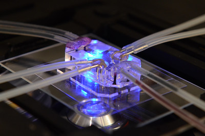
We have an interdisciplinary program with biomaterials, clinicians, surgeons, electrical engineers and chemical engineers to 3D fabricate cochleas with microanatomy similar to living cochleas, embedded with sensors that can sense current spread from cochlear implants, or ion gradients from various inner ear cell types that can generate them. Our goal is to develop these types of constructs, seed them with 3D cultures of various  inner ear cell types and examine how cochlear implants can activate auditory neurones, or how regeneration or pharmacologic support for hearing loss can be developed.  his is in order to develop the next generation of inner ear hearing loss therapies.

230
Category:
Biomedical Engineering & Biophysics
Project:

Develop Implantable BIOsensors for the detection of small METAbolites in the inflamed brain

Project Listed Date:
Institute or Center:
N/A
NIH Mentor:
N/A
University:
Cambridge
Project Details:

The project aims to develop and exploit high transconductance organic electrochemical transistor-based bio-sensors and ultra-low power thin-film electronics as emerging ICT tools with perfect fit to the targeted application domain. The proposed sensors and interfaces will provide unprecedented ability to detect and monitor small metabolites both in vitro and in vivo, to map immunometabolism of organs and tissues, and to test new drugs in situ.

222
Category:
Biomedical Engineering & Biophysics
Project:

Invent and implement new radioactive probes for imaging specific molecular targets 

Project Listed Date:
Institute or Center:
National Institute of Mental Health (NIMH)
NIH Mentor:

Dr. Victor Pike

University:
Cambridge
Project Details:

Invent and implement new radioactive probes for imaging specific molecular targets in animal and human brain with positron emission tomography

195
Category:
Biomedical Engineering & Biophysics
Project:

Invent and implement new radioactive probes for imaging specific molecular targets

Project Listed Date:
Institute or Center:
National Institute of Mental Health (NIMH)
NIH Mentor:

Dr. Victor Pike

University:
Oxford
Project Details:

Invent and implement new radioactive probes for imaging specific molecular targets in animal and human brain with positron emission tomography

175
Category:
Biomedical Engineering & Biophysics
Project:

Identifying mechanisms by which endothelial cells sense and respond to blood flow

Project Listed Date:
Institute or Center:
N/A
NIH Mentor:
N/A
UK Mentor:

Prof. Ellie Tzima

University:
Oxford
Project Details:

Forces are important in the cardiovascular system, acting as regulators of vascular physiology and pathology. Vascular endothelial cells are constantly exposed to mechanical forces, such as shear stress, due to the flowing blood. Patterns of blood flow depend on blood vessel geometry and type and can range from uniform blood flow (which is protective) to disturbed blood flow (which is pathologic). Although we know that endothelial cells can sense and respond differently to different types of flow, the mechanisms by which they sense and respond to blood flow remain a mystery. Our laboratory has pioneered the studies of endothelial mechano-sensing and has championed the use of a multi-disciplinary approach to this scientific problem. The focus of the proposed studentship is to identify mechanisms by which endothelial cells sense and respond to blood flow.  The student will have the opportunity to be exposed to a wide range of techniques based on the student’s individual interests that include: i) use of imaging and genetic approaches to characterize how mechano-sensing affects disease initiation and progression ; (2) applying high throughput RNA sequencing and proteomics approaches to globally dissect steps involved in disease etiology; 3) use of bioinformatics and biochemical experimental approaches to understand the role of blood flow forces in cardiovascular disease.

170
Category:
Biomedical Engineering & Biophysics
Project:

Ion Channel Gating and Biophysics

Project Listed Date:
Institute or Center:
N/A
NIH Mentor:
N/A
University:
Oxford
Project Details:

We have been studying the mechanism of gating in the Two-Pore Domain (K2P) family of K+ channels and the way in which their gating can be regulated by lipids and small molecules.  It is now clear these channels use a variety of structural mechanisms to open and close their pores, including changes within the selectivity filter itself.  This mechanism of filter gating is also known to occur in other members of this superfamily of tetrameric cation channels including the BK Calcium-activated K+ channel and Cyclic Nucleotide Gated (CNG) channels.  In the proposed project the student would have the opportunity to combine multiple different biophysical, computational, and functional approaches to investigate the structural mechanisms of filter gating in these channels and investigate what properties might be common amongst these channels.

102
Category:
Biomedical Engineering & Biophysics
Project:

Myosin characterization using light microscopy

Project Listed Date:
Institute or Center:
National Heart, Lung, and Blood Institute (NHLBI)
NIH Mentor:

Dr. James Sellers

University:
Oxford
Project Details:

This project would be to extend a long standing and productive collaboration between the Kukura lab at Oxford and the Sellers lab at NIH.  The specialty of the Kukura lab is in the development of light microscopic approaches to study novel biological processes with high temporal and spatial resolution.  The strength of the Sellers lab is in the production and characterization of myosins using various biochemical and biophysical approaches.

Back to Top