header-bg

Research Opportunities

Background Header
Image
Prospective Students

The goal of the NIH Oxford-Cambridge (OxCam) Scholars Program is to create, foster, and advance unique and collaborative research opportunities between NIH laboratories and laboratories at the University of Oxford or the University of Cambridge. Each OxCam Scholar develops a collaborative research project that will constitute his/her doctoral training. Each Scholar also select two mentors – one at the NIH and one in the UK – who work together to guide the Scholar throughout the research endeavor.

Students may select from two categories of projects: Self-designed or Prearranged. OxCam Scholars may create a self-designed project, which enables students to develop a collaborative project tailored to his/her specific scientific interests by selecting one NIH mentor and one UK mentor with expertise in the desired research area(s). Alternatively, students may select a prearranged project provided by NIH and/or UK Investigator(s) willing to mentor an OxCam Scholar in their lab.

Self-designed Projects 
Students may create a novel (or de novo) project based on their unique research interests. Students have the freedom to contact any PI at NIH or at Oxford or Cambridge to build a collaboration from scratch. The NIH Intramural Research Program (IRP) represents a community of approximately 1,200 tenured and tenure-track investigators providing a wealth of opportunity to explore a wide variety of research interests. Students may visit https://irp.nih.gov to identify NIH PIs performing research in the area of interest. For additional tips on choosing a mentor, please visit our Training Plan.

Prearranged Projects
Investigators at NIH or at Oxford or Cambridge have voluntarily offered collaborative project ideas for NIH OxCam Scholars. These projects are provided below and categorized by research area, NIH Institute/Center, and University. In some cases, a full collaboration with two mentors is already in place. In other instances, only one PI is identified, which allows the student to select a second mentor to complete the collaboration. Please note that prearranged project offerings are continuously updated throughout the year and are subject to change.

6 Search Results

CAPTCHA
719
Category:
Structural Biology
Project:

Structural mechanisms of TCR recognition of cancer antigen pMHC 

Project Listed Date:
Institute or Center:
N/A
NIH Mentor:
N/A
UK Mentor:

Prof. Peijun Zhang

University:
Oxford
Project Details:

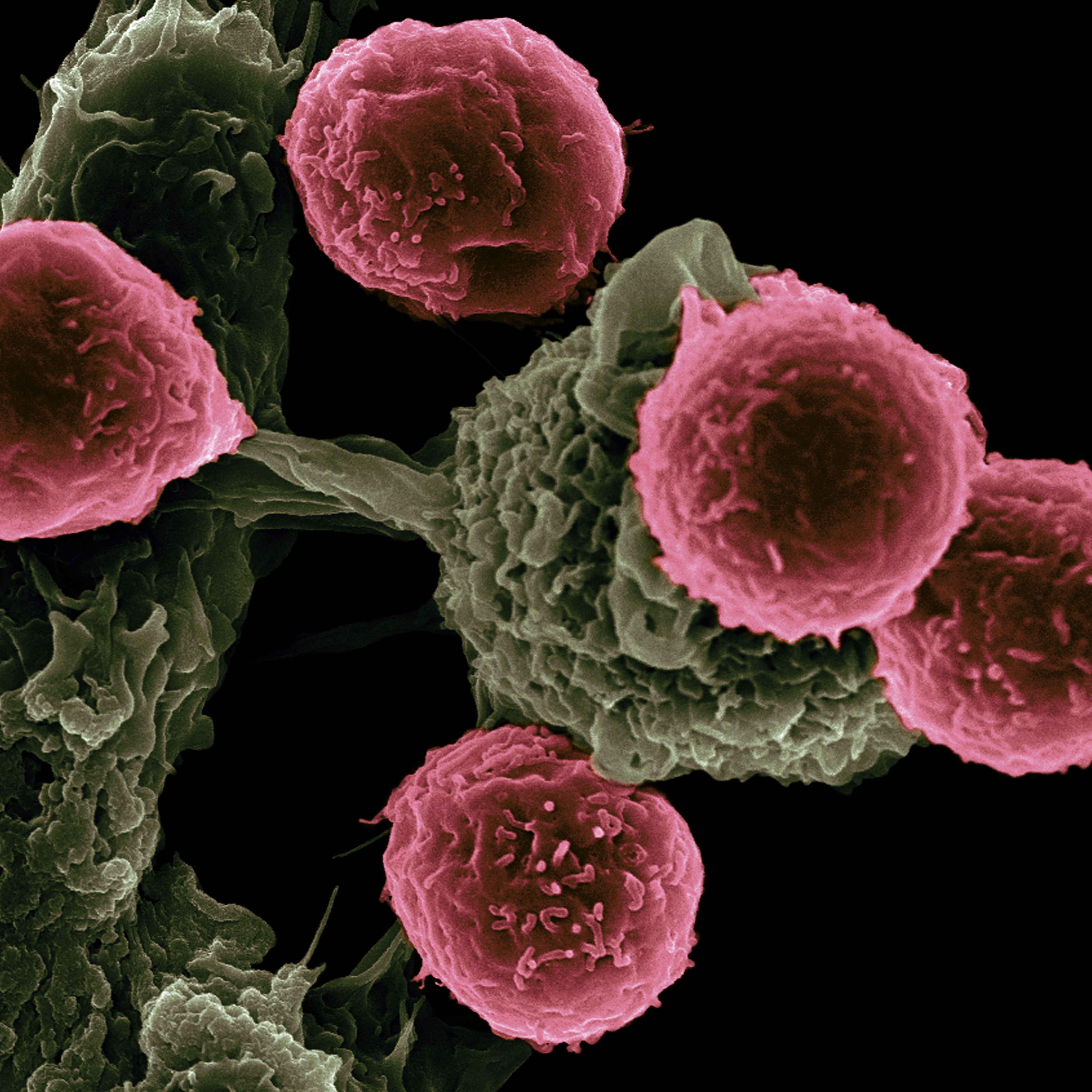
Immune surveillance plays a formidable role in eliminating cancer cells. At the centre of this process lies an interaction between the T cell receptor (TCR) in T cells and peptides presented by the major histocompatibility complex (pMHC). The biochemical and structural properties that underpin the TCR/pMHC interaction dictate the fate of infected or cancer cells. However, the rules that govern TCR specificity and sensitivity remain unknown. 

We aim to apply cryoEM and cryo-electron tomography (cryoET) including in situ structural studies to determine the structural fingerprints associated with productive TCR/pMHC interactions. We have recently made crucial technical advances in determining the structures of native unmodified TCR/pMHC complexes derived from SARS-CoV-2 infection, which had been intractable due to their intrinsic flexibility and innate low affinity. A robust cryoEM structure pipeline has thus been established for elucidating tumour-reactive TCRs in complex with natural cancer pMHCs. We will examine conserved features and differences among complexes to understand the observed specificity, sensitivity and binding affinity of TCRs recognising cancer antigens. We will also determine the complete, native TCR/CD3 and pMHC complexes from opposing membranes as they are in the immunological synapse formed between the T cell and target cancer cell using in situ cryoET.

These structures will reveal mechanisms underpinning the specificity and affinity for each TCR/pMHC pair and common shared interfaces. Such structural information will make critical contributions to the selection of TCRs for the deployment of robust and safe TCR-based immunotherapies

718
Category:
Structural Biology
Project:

Structural basis of HIV-1 nuclear import by in situ cryo-tomography and correlative microscopy

Project Listed Date:
Institute or Center:
N/A
NIH Mentor:
N/A
UK Mentor:

Prof. Peijun Zhang

University:
Oxford
Project Details:

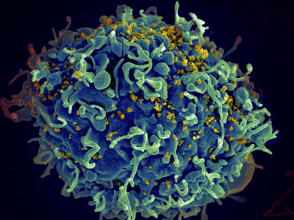
Human immunodeficiency virus type 1 (HIV-1) is the causative agent behind acquired immunodeficiency syndrome (AIDS) that currently has no cure or vaccine. While antiviral treatments are effective, the rise of drug-resistant strains has become a growing concern. HIV-1 primarily infects the immune system, targeting CD4+ T cells and macrophages and is a lentivirus known to be able to infect non-dividing cells, requiring it to exploit nuclear import mechanisms. This process is dependent on the viral capsid. The HIV capsid is a conical structure that houses the genomic material of the virus. It needs to be metastable in order to be protective while allowing timely disassembly (termed uncoating) to release its genome. The dynamics of the capsid nuclear import and uncoating are still unknown and are modulated by host dependency and restriction factors.

We aim to apply multi-imaging modalities to investigate uncoating and nuclear import of HIV. These will include super-resolution fluorescence microscopy (including the newest MINFLUX technology), Focused Ion Beam and Scanning electron microscopy (cryoFIB/SEM), cryo-electron microscopy and cryo-electron tomography (cryoEM/ET). The viral core and host factors will be fluorescently tagged, and infection will be monitored from viral attachment to nuclear import. The sample will be cryo-preserved and imaged by cryoEM/ET and cryoFIB/SEM. The combination of these imaging techniques, paired with molecular biology and virology tools, will yield unparalleled knowledge of the HIV infection process within the native cells, providing the framework for development of novel therapeutics targeting HIV infection in the future.

645
Category:
Structural Biology
Project:

Structural Analysis of Centrosome Biogenesis

Project Listed Date:
Institute or Center:
National Cancer Institute (NCI)
NIH Mentor:

Dr. Susan Lea

UK Mentor:

Prof. Jordan Raff

University:
Oxford
Project Details:

Most eukaryotic cells are born with a single centrosome that plays an important part in many aspects of cellular organization. Centrosomes are an excellent model for studying organelle biogenesis because, just like the DNA, they duplicate precisely once during each cell cycle. In the early Drosophila embryo, hundreds of centrosomes synchronously assemble every few minutes as the embryos rapidly progress through repeated cycles of division. 

Although centrosomes are complex nanomachines comprising >400 proteins, only ~10 proteins are absolutely essential for centrosome biogenesis. Thus, to understand the principles that allow these embryos to coordinately assemble so many centrosomes at the right time, in the right place, and then grow them to the right size, we need to understand how these proteins interact with each other, and how these interactions are regulated. This project involves using protein prediction software (e.g. AlphaFold2, Rosetta) to identify putative interactions and then using various approaches (biochemistry, CryoEM, in vitro reconstitution) to validate them (NIH). Once validated, the functional significance of these interactions will be tested using live-cell imaging in the early Drosophila embryo (Oxford). See publications below for two examples of previous collaborations between the NIH and Oxford groups.

Feng et al., Structural basis for mitotic centrosome assembly in flies. Cell, 2017.

Conduit et al., The centrosome-specific phosphorylation of Cnn by Polo/PLK1 drives Cnn scaffold assembly and centrosome maturation. Dev. Cell., 2014.

642
Category:
Structural Biology
Project:

Structure-function studies of mechanisms underlying the modulation of WNT signalling by R-spondins

Project Listed Date:
Institute or Center:
National Cancer Institute (NCI)
University:
Oxford
Project Details:

The WNT signalling pathway regulates patterning and morphogenesis during embryonic development and promotes the renewal of stem cells to maintain tissue homeostasis in adults. Aberrant WNT signalling also drives many types of cancer. Some WNT responses in vertebrates depend on a second signal provided by the R-spondin family of four secreted proteins, RSPO1-4. RSPOs markedly amplify target cell sensitivity to WNT ligands by neutralizing two transmembrane ubiquitin ligases, ZNRF3 and RNF43, which reduce the cell surface levels of WNT receptors. RSPOs can simultaneously engage ZNRF3/RNF43 and the 7-pass transmembrane receptors LGR4, 5 or 6 to trigger the clearance of ZNRF3/RNF43 from the cell surface, followed by lysosomal degradation. RSPO2 and RSPO3 can also engage heparan sulfate proteoglycans (HSPGs) such as glypicans or syndecans to promote ZNRF3/RNF43 clearance in the presence or absence of LGRs. In both cases, ZNRF3/RNF43 clearance results in increased WNT receptor levels at the cell surface and higher sensitivity to WNT ligands.

The molecular mechanism whereby binding of RSPOs to their LGR and/or HSPG receptors and to their ZNRF3/RNF43 effectors promotes the clearance of the trimeric or tetrameric complexes from the cell surface has remained elusive. In this project, students will combine genetic, cell biological, biochemical, biophysical and structural approaches in the laboratories of Dr. Jones at Oxford University and Dr. Lebensohn at the National Cancer Institute to elucidate the underlying mechanisms.

431
Category:
Structural Biology
Project:

Self-assembling multi-functional biomolecular condensates for targeted degradation of disease-associated proteins 

Project Listed Date:
Institute or Center:
N/A
NIH Mentor:
N/A
University:
Cambridge
Project Details:

We are developing artificial multi-valent proteins capable of liquid-liquid phase separation (LLPS) with the aim of building multi-functional biomolecular condensates and thereby harnessing specific cellular enzymes to target disease-associated proteins for destruction. We propose to design condensates that contain a class of proteins known as tandem-repeat proteins (RPs). We have shown that RPs are strikingly amenable to rational design and can be engineered to simultaneously bind multiple proteins, bringing them into specific spatial proximity in such a way as to enable a chemical modification of the target protein. The rational design of LLPS systems capable of selectively recruiting client proteins into them to drive specific biological reactions would enable both a deeper understanding of the role of biomolecular condensates in nature as well as the exploitation of their remarkable physico-chemical properties for therapeutic effect. 

Key areas of interest include: 
 

  1. Understanding the molecular grammar of protein phase separation to define rules for creating designer LLPS systems. 
  1. Developing novel hetero-bifunctional phase-separating proteins to recruit disease-associated targets to the protein degradation machinery. 
  1. Translating the designed LLPS proteins into biomolecular condensates in the cell capable of enhancing targeted protein degradation. 
414
Category:
Structural Biology
Project:

Structure-function studies of bacterial cell envelope machines involved in pathogenesis

Project Listed Date:
Institute or Center:
National Cancer Institute (NCI)
NIH Mentor:

Dr. Susan Lea

UK Mentor:

Prof. Ben Berks

University:
Oxford
Project Details:

The bacterial cell envelope comprises the cell wall and either one or two membranes. The cell envelope is of major interest in infection biology because it is the site at which pathogenic bacteria interact with their host organism. In particular, bacterial virulence proteins must be transported across the cell envelope to affect the host.

We aim to understand the molecular mechanisms by which proteins, nucleic acids, and mechanical force are transferred across and along the cell envelope in processes of biomedical importance. The project is to undertake structure-led studies of  the dedicated nanomachines that carry out these processes. The NCI part of the collaboration will involve structural analysis, primarily by cryoEM. The Oxford part of the collaboration will concentrate on complementary mechanistic work using biochemical, genetic, and live cell single molecule imaging methods. Our groups  have collaborated for more than 15 years on projects including the transport of folded proteins across the bacterial inner and outer membranes (Tat protein transport system and Type 9 Secretion System), lipoprotein export, DNA transport during horizontal gene transfer (conjugation and natural competence), and gliding motility.

Back to Top