header-bg

Research Opportunities

Background Header
Image
Prospective Students

The goal of the NIH Oxford-Cambridge (OxCam) Scholars Program is to create, foster, and advance unique and collaborative research opportunities between NIH laboratories and laboratories at the University of Oxford or the University of Cambridge. Each OxCam Scholar develops a collaborative research project that will constitute his/her doctoral training. Each Scholar also select two mentors – one at the NIH and one in the UK – who work together to guide the Scholar throughout the research endeavor.

Students may select from two categories of projects: Self-designed or Prearranged. OxCam Scholars may create a self-designed project, which enables students to develop a collaborative project tailored to his/her specific scientific interests by selecting one NIH mentor and one UK mentor with expertise in the desired research area(s). Alternatively, students may select a prearranged project provided by NIH and/or UK Investigator(s) willing to mentor an OxCam Scholar in their lab.

Self-designed Projects 
Students may create a novel (or de novo) project based on their unique research interests. Students have the freedom to contact any PI at NIH or at Oxford or Cambridge to build a collaboration from scratch. The NIH Intramural Research Program (IRP) represents a community of approximately 1,200 tenured and tenure-track investigators providing a wealth of opportunity to explore a wide variety of research interests. Students may visit https://irp.nih.gov to identify NIH PIs performing research in the area of interest. For additional tips on choosing a mentor, please visit our Training Plan.

Prearranged Projects
Investigators at NIH or at Oxford or Cambridge have voluntarily offered collaborative project ideas for NIH OxCam Scholars. These projects are provided below and categorized by research area, NIH Institute/Center, and University. In some cases, a full collaboration with two mentors is already in place. In other instances, only one PI is identified, which allows the student to select a second mentor to complete the collaboration. Please note that prearranged project offerings are continuously updated throughout the year and are subject to change.

241 Search Results

CAPTCHA
455
Category:
Computational Biology
Project:

Understanding the clonality of drug resistance in cancer

Project Listed Date:
Institute or Center:
N/A
NIH Mentor:
N/A
UK Mentor:

Prof. Adam Cribbs

University:
Oxford
Project Details:

We are interested in understanding the clonality of drug resistance in cancer (primarily in a haematological cancer called Multiple Myeloma (MM)). To achieve our goals, we have developed state of the art long-read single-cell sequencing approaches (termed scCOLOR-seq) that allow us to measure single clones within patient samples. This method allows for the simultaneous measurement of gene expression, exon mutations, exon SNPs and translocations. We apply this technology and develop cutting edge computational analysis solutions to understand the relationship between clonality and drug resistance in oncology. Furthermore, our lab is also working as part of the Human Cell Atlas (HCA) project and we have several international collaborations with immunologists, cancer biologists to support our work.

The aim of this project is to develop computational analysis strategies aimed at better defining specific MM clones within patients that are resistant to first line therapeutics. The work will involve combining long-read (Oxford Nanopore Technology) multi-modal datasets and performing machine learning approaches to better define high risk patients. This work is important to better understand drug resistance mechanisms in MM and identify patients that may respond less well to therapy.

453
Category:
Project:

Investigating mechanisms of ER-associated degradation (ERAD) in human disease

Project Listed Date:
Institute or Center:
National Cancer Institute (NCI)
NIH Mentor:

Dr. Susan Lea

University:
Oxford
Project Details:

Accumulation of misfolded proteins and aberrant protein aggregates are hallmarks of a wide range of pathologies such as neurodegenerative diseases and cancer. Under normal conditions, these potentially toxic protein species are kept at low levels due to a variety of quality control mechanisms that detect and selectively promote their degradation. Our lab investigates these protein quality control processes with a particular focus on ER-associated degradation (ERAD), that looks after membrane and secreted proteins. The ERAD pathway is evolutionarily conserved and in mammals, targets thousands of proteins influencing a wide range of cellular processes, from lipid homeostasis and stress responses to cell signaling and communication.

We investigate the mechanisms of ERAD using multidisciplinary approaches both in human and yeast cells. Using CRISPR-based genome-wide genetic screens and light microscopy experiments we identify and characterize molecular components involved in the degradation of disease-relevant toxic proteins. In parallel, we use biochemical tools to dissect mechanistically the various steps of the ERAD pathways. In this collaborative project with the Lea lab we will use structural approaches such as cryo-electron microscopy to gain insight into the molecular mechanisms of ERAD.

These studies, by providing mechanistic understanding of the ERAD process, may shed light on human diseases impacting ER function and may ultimately contribute to better therapeutics. 

452
Category:
Neuroscience
Project:

Combining neuroimaging and neurophysiology to understand the nature of residual vision across species following damage to primary visual cortex

Project Listed Date:
Institute or Center:
National Institute of Mental Health (NIMH)
NIH Mentor:

Dr. David Leopold

UK Mentor:

Prof. Holly Bridge

University:
Oxford
Project Details:

The laboratory of Prof Bridge in Oxford focusses on understanding the pathways in the human visual system that can process residual vision after someone has had a stroke that affects the primary visual cortex.

The laboratory of Prof Leopold combines neuroimaging, behaviour and neurophysiology in a non-human primate model to better understand computation in the visual system, particularly relating to conscious perception.

The proposed PhD project would have 3 main objectives:

  1. Quantitatively compare changes in retinotopic maps and population receptive fields in humans and non-human primates with damage to primary visual cortex.
  2. Determine the visual pathways in the two species that are necessary and/or sufficient to provide residual vision within the blind region of the visual field.
  3. Investigate the neural changes that occur as a result of visual training following the damage to the visual system in order to inform rehabilitation programmes for people who have suffered a stroke to the visual system.

    During the training programme, the student would have the opportunity to learn about multi-modal human neuroimaging approaches applied to both the healthy and the damaged visual system. This would be complemented by training in both neuroimaging and neurophysiology in the non-human primate.
451
Category:
Neuroscience
Project:

Investigating peripheral sensory neuron circuits in health and disease.

Project Listed Date:
Institute or Center:
National Institute of Health (NIH)
NIH Mentor:

Dr. Alex Chesler 

University:
Oxford
Project Details:

Our aim is to understand the sensory circuits that govern normal (protective) touch and pain, and how following injury or disease to the nervous system, pain can become chronic (neuropathic pain). Sensory neurons are heterogeneous neurons that innervate sensory targets (such as the skin), and extend central terminals which enter the dorsal horn of the spinal cord. We are interested in the role of molecularly defined subpopulations of sensory neurons, and how they contribute to the development and maintenance of neuropathic pain. We utilise chemogenetic and optogenetic strategies to selectively silence or activate sensory neuron function in mice. Combining this with behavioural paradigms to assess evoked and spontaneous pain, allows us to identify key populations of sensory neurons in pain processing, with the hope of identifying new druggable targets for future development. Further validation is undertaken in human iPSC derived sensory neurons. Students will learn and use a wide range of techniques including (but not limited to); Chemogenetics, optogenetics, viral vectors, mouse transgenics, animal behaviour, electrophysiology and calcium imaging.

450
Category:
Stem Cell Biology
Project:

Elucidating disease mechanisms in cerebellar ataxia using stem cell technology

Project Listed Date:
Institute or Center:
National Heart, Lung, and Blood Institute (NHLBI)
NIH Mentor:

Dr. John A. Hammer 

University:
Oxford
Project Details:

The spinocerebellar ataxias (SCAs) are a complex group of neurodegenerative diseases that affect the cerebellum and result in the loss of motor coordination. No effective treatments exist for the SCAs, and there is a pressing need for better models in which to study the underlying disease-causing mechanisms and to identify potential therapies.

The aim of this project will be to develop novel stem cell-derived models to identify common pathological mechanisms in SCA that could be targeted therapeutically. The Becker group has identified several novel SCA mutations that highlight mGluR1-TRPC3-IP3R1 signaling as a key pathway affected in disease. Both research groups have developed complementary stem cell-derived and primary cerebellar models that provide unique systems to investigate the functional consequences of gene mutations affecting this pathway in the Purkinje cells, which are the neurons that are primarily affected in SCA.

The project will employ patient-derived induced pluripotent stem cells (iPSCs) as well as introduce gene mutations into iPSCs using CRISPR gene editing technology. Pluripotent stem cells will be differentiated into cerebellar neurons and three-dimensional organoids and deeply phenotyped using a combination of functional experiments including calcium imaging, super-resolution imaging, and morphological analyses. In addition, functional analyses will be carried out in primary Purkinje cells. Identified disease phenotypes will subsequently be screened for potential therapeutics.


Becker Group website: https://www.ndcn.ox.ac.uk/research/cerebellar-disease-group

Hammer Group website: https://irp.nih.gov/pi/john-hammer

449
Category:
Immunology
Project:

Investigating B and T cell populations in health and disease

Project Listed Date:
Institute or Center:
N/A
NIH Mentor:
N/A
University:
Oxford
Project Details:

How are different B cell populations developmentally linked in human health and disease?

We are investigating the generation, function and plasticity of B cell populations in human health. In particular, we are interested in how different lymphocyte subsets are developmentally linked and differences in function, and therefore providing a platform to understand how B cell fate may be different in human disease. We are defining how B cells select a particular developmental pathway, and will use this information to develop methods for modulating B cell function as potential therapeutic approaches.

How can B and T cells may be therapeutically modulated across cancers and autoimmune diseases?

There is accumulating evidence for the role of both T and B cells in modulating immune responses to both solid tumours and haematological malignancies. We are investigating the contributions, function and heterogeneity of B and T cells on the immune responses to tumours and their potential role in cancer detection and treatment. We are determining the nature of B and T cell immuno-surveillance, regulation and activation across cancers and autoimmune diseases, as well as the immunological features associated with better prognosis and immunomodulation. With this, we aim to highlight novel therapeutic avenues. Our lab is affiliated with the Oxford Cancer Centre (https://www.cancer.ox.ac.uk/research/research-themes/developments-in-immuno-oncology) and non-cancer clinicians, with strong clinical links to a wide range of hard-to-treat diseases.

What is the effect of genetic and environmental variation on B and T cell fate?

Immunological health relies on a balance between the ability to mount an immune response against potential pathogens and tolerance to self. B and T cells are key to the immune response by producing antibodies and cytotoxic T cells. B/T cell clones selectively expand following antigen recognition by B and T cell receptors (BCR and TCR) respectively. BCRs are the membrane-form of antibodies and are generated through DNA recombination resulting in the potential to recognise a vast array of pathogens. Defects in the ability to mount effective B cell or T cell responses have been implicated in infectious susceptibility, impaired surveillance of cancer and immunodeficiencies, whereas a breakdown of immunological tolerance has been attributed to autoimmune diseases such as through autoantibody production and reduced numbers of regulatory B/T cells. Through integrating genomics, bulk and single-cell transcriptomics, and metabolomics data, serological, B /T cell repertoire and viromics datasets we will investigate the effect of both genetic variation and environmental factors on B cell fate, regulation, and the relationship to disease susceptibility.

448
Category:
Immunology
Project:

Defining the role of shared T-cell receptor clonotypes in SARS-CoV-2 infection

Project Listed Date:
Institute or Center:
N/A
NIH Mentor:
N/A
UK Mentor:

Prof. Peijun Zhang

University:
Oxford
Project Details:

T cells are a major component of adaptive immune response, continuously screening lymphoid tissues for antigen peptides presented by major histocompatibility complex (peptide/MHC or pMHC)3. These antigen peptides are recognized by T-cell receptors (TCRs). Thymocytes with a low-affinity TCRs mature into T cells and enter the lymphoid organs, where they are exposed to foreign antigen peptides by MHC molecules from antigen-presenting cells including macrophages, dendritic cells, and B-cells, during infection. When the T-cell receptors bind to antigenic peptide, T-cells are activated and undergo clonal expansion, resulting in immune response. CD8+ T cells play an important for immune response and viral clearing, but their role in protection and pathogenesis of SARS-CoV-2 remains poorly understood4. In addition to extensively studied spike protein, open reading frame 3a (ORF3a), a highly conserved protein within the Betacoronavirus subgenus, has been considered as a potential target for vaccines or therapeutics, with deletion of ORF3a resulting in decreased viral titer and morbidity. We have identified shared CD8+ T cell clonotypes responding to a ORF3a in COVID-19 infections.  Importantly, shared clonotypes in severe COVID-19 infections provides a target for development of novel antiviral immunotherapies. The aim of this project is to analyse shared TCR clonotypes in ORF3a recognition and provide structural basis for the recognition of ORF3a-pMHC complex by T-cell receptors.

447
Category:
Cell Biology
Project:

Analysis of APP and Tau function in exosome and supermere secretion and signalling

Project Listed Date:
Institute or Center:
N/A
NIH Mentor:
N/A
UK Mentor:

Prof. Clive Wilson

University:
Oxford
Project Details:

Aggregations of the cytoskeletal protein Tau into neurofibrillary tangles and cleavage products of the Amyloid Precursor Protein (APP) into amyloid plaques are strongly associated with the progression of Alzheimer’s Disease (AD). However, it remains unclear whether these aggregates initiate and cause disease or are primarily by-products of more fundamental defects. Furthermore, the physiological and cellular roles of Tau and APP, which might be disrupted in AD, are not well understood.

We have developed a new cell model in the fruit fly, Drosophila melanogaster, to study the normal functions of fly APP and Tau. Remarkably, we find that they are both involved in the biogenesis and secretion of two multimolecular signalling complexes, which are formed in endosomes: exosomes, which are small secreted vesicles, and supermeres, newly identified aggregates of protein and RNA. Their secretion is disrupted when pathological versions of Tau or cleaved APP are expressed in these cells. Using the fly model, we have already identified multiple additional regulators of these APP- and Tau-dependent processes, some of which are implicated in other neurodegenerative diseases. This project will employ neuronal and cancer cell lines to investigate which of these mechanisms are conserved in human cells and how they affect both exosome and supermere signalling. Again, informed by genetic screens in flies, we will then test whether Tau- and APP-induced defects in secretion are suppressed by genetic manipulations of other genes, which might provide new therapeutic targets going forward.

446
Category:
Biomedical Engineering & Biophysics
Project:

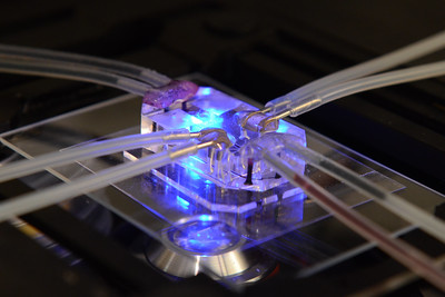
Development of a high throughput 3D-microfluidics-based blood-brain barrier assay system as an in vitro model for ultrasound-mediated delivery of drugs into the brain

Project Listed Date:
Institute or Center:
N/A
NIH Mentor:

Dr. Marc Ferrer 

University:
Oxford
Project Details:

More than 90% of the drugs that enter clinical trials fail because of lack of efficacy or unexpected toxicity. This high failure rate has been partly attributed to the use of in vitro cellular assays and animal models that do not reproduce human physiology and pathology during preclinical drug development. This gap in clinical predictability in drug development is especially severe for CNS disorders, in which many therapeutics must cross the blood-brain barrier (BBB) to be effective. There has been a steady development and evaluation of drug delivery mechanisms that bypass the BBB to reach the target site in the CNS, and ultrasound (US)-mediated therapeutics extravasation has been one of such tools. However, while there have been several in vivo animal and clinical studies done on the focused US-guided BBB disruption, there have not yet been an established in vitro model in which US has been utilized to understand the BBB tight junction disruption and penetration. This project seeks to develop a microfluidics-based BBB model in order to: (1) investigate brain endothelial cell behaviour in microenvironment when stimulated by US and microbubbles; (2) establish whether US provides a means to enhance drug delivery through the BBB using the in vitro microfluidics-based assay platform; (3) investigate the stimulation of any inflammatory responses arising from ultrasound/microbubble exposure (in collaboration with NCI, Frank Lab). The work will contribute to greater understanding of interactions between US-mediated microbubble disruption of the BBB, especially benefitting the physicians working to utilize the technique in clinical settings and the researchers developing microbubbles to establish and optimize BBB drug delivery during the drug R&D pipeline.

445
Category:
Microbiology and Infectious Disease
Project:

Characterising antibiotic-induced collateral damage in the gut microbiome

Project Listed Date:
Institute or Center:
N/A
NIH Mentor:
N/A
UK Mentor:

Prof. Mathew Stracy

University:
Oxford
Project Details:

Antibiotics help clear an ongoing infection, but they can also cause significant collateral damage: they select for drug-resistant strains and cause dysbiosis to the commensal gut microbiota. This antibiotic-induced selection for resistance within the microbiome can facilitate the spread of resistant pathogens to extra-intestinal infections and to other patients (see our recent work: Stracy et al. Science. 2022, 375 (6583), 889-894).

This project will aim to understanding how antibiotics cause collateral damage to the microbiota leading to subsequent resistant infections. This will involve experiments with synthetic and human-derived microbial communities as well as developing microscopy methods to understand the effect of antibiotics on the micro-scale biogeography of the microbiota. The aim is to characterise the key factors that determine how antibiotics affect a patient’s resident microbial population and cause resistant opportunistic pathogens to spread. This will be used to help develop new ways to minimize the spread of antibiotic resistance both within and between patients. The project would suit applicants with a strong background in microbiology and keen interest in developing new ways to combat antibiotic resistance.

444
Category:
Cell Biology
Project:

Identifying novel targets to treat and prevent arterial disease

Project Listed Date:
Institute or Center:
N/A
NIH Mentor:
N/A
UK Mentor:

Prof. Nicola Smart

University:
Oxford
Project Details:

Progression of arterial disease is critically determined by the response of smooth muscle cells (SMCs) within the medial layer. In their fully differentiated, contractile state, SMCs confer stability and regulate vascular tone. However, disease induces a ‘contractile-synthetic’ phenotypic switch which impairs function, leads to vascular stiffness and exacerbates inflammation, to promote atherosclerosis and susceptibility to abdominal aortic aneurysm (AAA) (1). In animal studies, we have identified candidate pathways, based on knowledge of embryonic SMC differentiation, with the potential to protect against AAA1 and atherosclerosis (2) by preserving contractile SMC phenotype. However, the low success rate in translation from animal studies to the clinic highlights the need to determine whether similar mechanisms serve to protect the human vasculature, and how they may be targeted to alleviate disease.

The aim of the project is to establish human-relevant SMC models in which to study phenotypic switching and disease: i)
a monoculture of human coronary artery SMCs; the simple monoculture model will permit evaluation of factors that directly impact SMC modulation, without confounding influences of other cell types.  ii) a more physiologically relevant model of hcSMCs co-cultured with coronary arterial endothelial cells. EC-SMC interaction crucially maintains vascular tone and functionality and dysregulation of this crosstalk underpins pathological remodelling e.g. in intimal hyperplasia. These models will then be used for high genetic and pharmacological screening to reveal disease-modifying targets. The most promising will be explored in murine disease models.


1). Munshaw et al. (2021) J Clin Invest 131.(10):e127884.

2). Munshaw, Redpath, Pike & Smart. bioRxiv, 2021.2011.2030.470548 (2021).

443
Category:
Developmental Biology
Project:

Role of the cell cycle in controlling epicardial contribution to the developing and regenerating heart

Project Listed Date:
Institute or Center:
N/A
NIH Mentor:
N/A
UK Mentor:

Prof. Nicola Smart

University:
Oxford
Project Details:

Our group investigates embryonic mechanisms of cardiovascular development, to inform cardiac regenerative strategies, through reactivating of developmental processes in the adult heart1. The epicardium plays a crucial role in the embryo, to stimulate growth of the coronary vasculature and maturation of the myocardium (1). A key first step is epithelial to mesenchymal transition (EMT) to yield migratory cells which invade the myocardium and secrete potent paracrine factors. The adult mammalian epicardium is reactivated in response to myocardial infarction and contributes to repair (2), albeit sub-optimally, a major limitation being the extent of endogenous EMT. Based on our knowledge of ‘optimal’ embryonic mechanisms, we seek to enhance epicardial-based regeneration of the injured adult heart. Our preliminary data suggest a novel cell cycle-dependent mechanism controlling EMT and differentiation of epicardial cells, in line with the emerging paradigm of cell cycle control of stem cell fate (3). We will explore this exciting hypothesis by assessing how pharmacological and genetic perturbation of the cell cycle impacts EMT and fate, using functional assays, candidate and unbiased (e.g. RNA-Seq) approaches in cell culture, primary explant and genetic mouse models.

1). Redpath and Smart (2020). Stem Cells Transl Med. doi.org/10.1002/sctm.20-0352.

2). Smart et al. (2011) Nature. 474(7353):640-4.

3). Pauklin & Vallier (2013) Cell 155, 135.

442
Category:
Neuroscience
Project:

Neurobiological substrates of volitional social learning and memory

Project Listed Date:
Institute or Center:
National Institute on Drug Abuse (NIDA)
UK Mentor:

Prof. David Dupret

University:
Oxford
Project Details:

All memories, including social memories, are encoded in neuronal ensembles. Neuronal ensembles are small populations of sparsely distributed neurons selected by specific stimuli. We recently developed and published a mouse model of volitional social learning using a custom-made apparatus. This model provides a unique opportunity to study the neurobiological substrates of volitional social learning and memory. Previous studies have shown that the hippocampus, in particular the CA2 region, is critical for encoding social memories. However, almost all prior studies on the neurobiological basis of mouse social interaction fail to account for the volitional aspect of social interaction. The project we propose entails a collaboration between our lab at University of Oxford and Dr. Hope’s lab at NIDA IRP to investigate the role of hippocampal neuronal ensembles in volitional social learning and memory. Dr. Hope and Dr. Ramsey have developed and implemented the volitional social learning task, and my lab regularly performs in vivo recordings in neuronal ensembles of the hippocampus.  Thus, we will co-mentor a graduate student through the NIH OxCam program to investigate activity in the hippocampus that encodes volitional social memories.

441
Category:
Neuroscience
Project:

Developing diagnostic methods that aid clinicians in early identification and differential diagnosis

Project Listed Date:
Institute or Center:
N/A
NIH Mentor:
N/A
University:
Oxford
Project Details:

To date, neurodegenerative diseases have no cure and the clinical diagnosis is very challenging due to considerable overlap in the pathology and clinical symptoms. Thus, there is a strong unmet need for objective and sensitive diagnostic methods that can aid clinicians in early identification and differential diagnosis.

We have developed seed amplification assay (SAA) that detects alpha-synuclein (aSyn) aggregation in CSF of patients with synucleinopathies with much higher sensitivity and specificity than previously possible. Our findings also show that aSyn SAA is able to distinguish between Parkinson’s disease and multiple system atrophy and identify patients at high risk with REM sleep behaviour disorder prior to their conversion. Here, we suggest to build a “multiplex” SAA to detect multiple aggregating proteins (aSyn, tau and TDP-43) and test its ability to stratify between dementias of different aetiologies. We are in an exceptional position to deliver this goal because we have the necessary technical knowledge to build robust SAAs combined with access to unique clinico-pathological cohorts with longitudinal CSF and donated brain, where our assay can be tested and validated.

This proof-of-concept data will be used to further evaluate early and accurate diagnosis in larger longitudinal cohort of patients with mild cognitive impairment (MCI) and dementia. The proposed work aims to accelerate clinical trials by providing a “personal protein signature” that can be targeted by therapies tailor-made for individual patients aiming to lower the burden of these specific proteins (e.g. cocktail of vaccinations) and superior means to measure the efficacy of such treatments.

440
Category:
Neuroscience
Project:

Investigating the role of neurotransmitters GABA and Acetylcholine in sensory processing

Project Listed Date:
Institute or Center:
N/A
NIH Mentor:
N/A
University:
Oxford
Project Details:

Parkinson’s disease is typically considered to impact motor functions. However, non-motor symptoms, such as visual hallucinations, increase disease burden. Developing therapies for hallucinations in Parkinson’s disease has been challenging, since we do not fully understand what causes them to occur. In the healthy brain, successfully interpreting what one sees involves different neurotransmitters like GABA and Acetylcholine; hence it is possible that pathological processes involving these brain chemicals relate to the generation of visual hallucinations.

The project will investigate the role of neurotransmitters GABA and Acetylcholine in sensory processing in the healthy and Parkinsonian human brain. It will combine pharmacological interventions with human neuroimaging (functional MRI, MR Spectroscopy) and non-invasive brain stimulation, to provide putative targets for therapeutic interventions to alleviate visual hallucinations in Parkinson’s disease.

Research in Oxford will take place at the Wellcome Centre for Integrative Neuroimaging (https://www.win.ox.ac.uk) and the MRC Brain Network Dynamics Unit (https://www.mrcbndu.ox.ac.uk), hosted by the Physiological Neuroimaging Group (https://www.ndcn.ox.ac.uk/research/physiological-neuroimaging-group).

Back to Top