header-bg

Research Opportunities

Background Header
Image
Prospective Students

The goal of the NIH Oxford-Cambridge (OxCam) Scholars Program is to create, foster, and advance unique and collaborative research opportunities between NIH laboratories and laboratories at the University of Oxford or the University of Cambridge. Each OxCam Scholar develops a collaborative research project that will constitute his/her doctoral training. Each Scholar also select two mentors – one at the NIH and one in the UK – who work together to guide the Scholar throughout the research endeavor.

Students may select from two categories of projects: Self-designed or Prearranged. OxCam Scholars may create a self-designed project, which enables students to develop a collaborative project tailored to his/her specific scientific interests by selecting one NIH mentor and one UK mentor with expertise in the desired research area(s). Alternatively, students may select a prearranged project provided by NIH and/or UK Investigator(s) willing to mentor an OxCam Scholar in their lab.

Self-designed Projects 
Students may create a novel (or de novo) project based on their unique research interests. Students have the freedom to contact any PI at NIH or at Oxford or Cambridge to build a collaboration from scratch. The NIH Intramural Research Program (IRP) represents a community of approximately 1,200 tenured and tenure-track investigators providing a wealth of opportunity to explore a wide variety of research interests. Students may visit https://irp.nih.gov to identify NIH PIs performing research in the area of interest. For additional tips on choosing a mentor, please visit our Training Plan.

Prearranged Projects
Investigators at NIH or at Oxford or Cambridge have voluntarily offered collaborative project ideas for NIH OxCam Scholars. These projects are provided below and categorized by research area, NIH Institute/Center, and University. In some cases, a full collaboration with two mentors is already in place. In other instances, only one PI is identified, which allows the student to select a second mentor to complete the collaboration. Please note that prearranged project offerings are continuously updated throughout the year and are subject to change.

241 Search Results

CAPTCHA
651
Category:
Systems Biology
Project:

Quantitative imaging and pooled CRISPRi screening of single cells to understand transcription factor signaling dynamics

Project Listed Date:
Institute or Center:
National Institute on Aging (NIA)
NIH Mentor:

Myong-Hee "Mia" Sung

UK Mentor:
N/A
University:
N/A
Project Details:

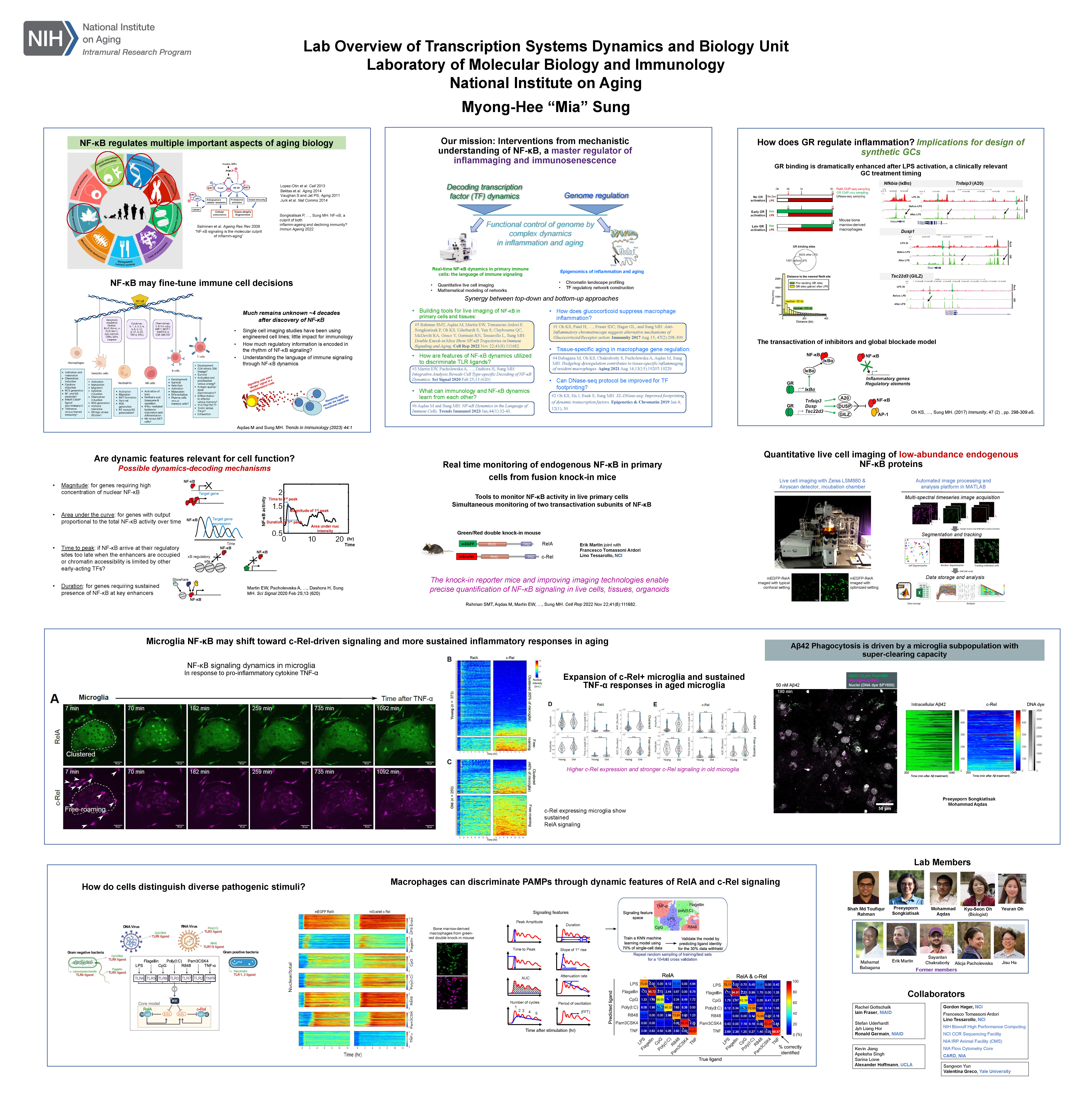
Quantitative imaging and pooled CRISPRi screening of single cells to understand transcription factor signaling dynamics. NF-κB is a master regulator of inflammation, immunity, and cell stress responses. The temporal dynamics of NF-κB signaling capture pathogen-specific information and govern the corresponding gene expression patterns. Recent studies have revealed that distinct NF-κB signaling profiles can lead to specific epigenetic modifications within potential enhancer regions of the genome, potentially establishing epigenetic memory for subsequent infections.

In this project, our goal is to investigate the impact of epigenetic perturbations on NF-κB signaling dynamics. We will utilize CRISPRi and a pool-genetic approach to systematically disrupt all potential enhancer regions around NF-κB-regulated genes. Additionally, we will conduct live-cell microscopy to quantitatively measure the resulting changes in NF-κB signaling dynamics. The insights gained from this study will illuminate the functions of the enhancer regions of NF-κB-regulated genes and will provide information on how tissue-specific NF-κB signaling is shaped by the epigenome, through the formation of epigenetic memories. The student will learn various interdisciplinary methods involving cell culture, quantitative microscopy, fluorescent reporter assays, automated single-cell analysis, molecular biology, and imaging data analysis.

650
Category:
Neuroscience
Project:

Mechanisms of perception and cognition 

Project Listed Date:
Institute or Center:
National Eye Institute (NEI)
NIH Mentor:

Dr. Bevil Conway

UK Mentor:
N/A
University:
N/A
Project Details:

Mechanisms of perception and cognition 
Section on Perception, Cognition, and Action, Laboratory of Sensorimotor Research (NEI/NIMH)

Students would have the option to work on any project in the lab, and to take it in new directions. Current projects in the lab aim to understand the normal brain processes by which physical signals that impinge on the sensory apparatus (eyes, ears) are transformed into perceptions, thoughts, and actions. Work in the lab has been especially invested in developing color as a model system. The advantage of color is that its physical basis (wavelength) is well characterized, yet these chromatic signals support not only low-level visual abilities such as color matching but also high-level cognitive processes such as categorization, memory, social cognition, and emotion. This variety of phenomena provides a rich opportunity for investigating the full scope of perceptual and cognitive computations that make human vision such an important source of information about the world. The lab uses many research techniques, including psychophysics and non-invasive brain imaging (MRI, MEG) in humans, along with fMRI-guided microelectrode recording, fMRI-guided pharmacological blockade, microstimulation, tract-tracing, and computational modeling in non-human primates (NHPs). Work in the lab is organized around Four broad approaches:

First, the use of MRI in humans and NHPs to investigate homologies of brain anatomy and function between these species, to support the applicability of neurophysiology from NHPs to the human case, and to test hypotheses about the fundamental organizational plan of the cerebral cortex in the primate.

Second, the use of well-controlled psychophysics (including longitudinal experiments) combined with microelectrode recording in NHPs to show on a mechanistic level how populations of neurons drive behaviors such as perceptual decisions, categorization, and concept formation and memory.

Third, comparative psychophysical studies in humans and NHPs, as part of a program of neuroethology to understand the relative computational goals of perception/cognition in different primate species. In addition to studies of vision, the lab conducts experiments using auditory and combined audio-visual stimuli, to understand common principles of sensory-cognitive information processing, and to determine how signals across the senses are integrated into a coherent experience.

Fourth, large-scale neurophysiological experiments combined with cutting-edge analysis methods including machine learning, to determine the mechanisms of high-acuity visual perception at the center of gaze. We have developed several eye-trackers that afford photo-receptor resolution, providing an unprecedented look at fine-scale spatial and chromatic processing of the foveal representation in primary visual cortex. 
 

649
Category:
Neuroscience
Project:

Investigating brain tumor signaling and development of therapeutics

Project Listed Date:
Institute or Center:
National Cancer Institute (NCI)
NIH Mentor:

Dr. Zhengping Zhuang

UK Mentor:

Dr. Harry Bulstrode and 
Dr. Justin Lathia (Cleveland Clinic)

University:
Cambridge
Project Details:

This collaboration will provide the opportunity to work on malignant brain tumors in the laboratories of these three investigators with complementary expertise. The Zhuang laboratory (NIH) has an interest in the investigation of hypoxia (HIF-2a) signaling and tumor development as well as the pathogenesis of brain tumors and development of therapeutics. The Bulstrode laboratory (Cambridge) has an interest in neural stem cell identity and microenvironment interactions in brain development and in brain tumors. The Lathia laboratory (Cleveland Clinic) has an interest in cancer stem cells, tumor microenvironment interactions, immune suppression, and sex differences in brain tumors. The prospective student will have access to mentorship, infrastructure, and resources across all three laboratories. 

646
Category:
Microbiology and Infectious Disease
Project:

Engineering the microbiota to reduce the spread of antibiotic resistance

Project Listed Date:
Institute or Center:
N/A
NIH Mentor:
N/A
UK Mentor:

Prof. Mathew Stracy

University:
Oxford
Project Details:

Antibiotics are crucial for the treatment of bacterial infections, but they can also cause significant collateral damage to the microbiota (de Nies et al. Nat Rev Microbiol. 2023). Antibiotics supress the growth of commensal microorganisms and at the same time select for drug-resistance. Individuals are often benignly colonized with resistant potential pathogens persisting at low levels within their microbiota, such as extra-intestinal pathogenic Escherichia coli (ExPEC). Antibiotics can lead to overgrowth of these resistant pathogens, facilitating their spread within and between individuals (Stracy et al. Science. 2022).Antibiotics can also lead to resistance genes spreading between bacteria within the microbiota through various horizontal gene transfer (HGT) mechanisms.

This overall aim of this project is to answer the important questions:

  1. What are the key factors that determine the level of antibiotic-induced resistance overgrowth/spread within the microbiota?
  2. How does antibiotic-induced pathogen overgrowth and level of HGT differ between individuals?
  3. Can we engineer the microbiota to prevent the spread of resistance?
     

To achieve this, we will use experimental approaches with synthetic and human-derived microbial communities as well as developing microscopy methods to understand the effect of antibiotics on the micro-scale biogeography of the microbiota. We will test approaches to modify the members of the microbiota to minimize antibiotic-induced pathogen overgrowth or HGT.

645
Category:
Structural Biology
Project:

Structural Analysis of Centrosome Biogenesis

Project Listed Date:
Institute or Center:
National Cancer Institute (NCI)
NIH Mentor:

Dr. Susan Lea

UK Mentor:

Prof. Jordan Raff

University:
Oxford
Project Details:

Most eukaryotic cells are born with a single centrosome that plays an important part in many aspects of cellular organization. Centrosomes are an excellent model for studying organelle biogenesis because, just like the DNA, they duplicate precisely once during each cell cycle. In the early Drosophila embryo, hundreds of centrosomes synchronously assemble every few minutes as the embryos rapidly progress through repeated cycles of division. 

Although centrosomes are complex nanomachines comprising >400 proteins, only ~10 proteins are absolutely essential for centrosome biogenesis. Thus, to understand the principles that allow these embryos to coordinately assemble so many centrosomes at the right time, in the right place, and then grow them to the right size, we need to understand how these proteins interact with each other, and how these interactions are regulated. This project involves using protein prediction software (e.g. AlphaFold2, Rosetta) to identify putative interactions and then using various approaches (biochemistry, CryoEM, in vitro reconstitution) to validate them (NIH). Once validated, the functional significance of these interactions will be tested using live-cell imaging in the early Drosophila embryo (Oxford). See publications below for two examples of previous collaborations between the NIH and Oxford groups.

Feng et al., Structural basis for mitotic centrosome assembly in flies. Cell, 2017.

Conduit et al., The centrosome-specific phosphorylation of Cnn by Polo/PLK1 drives Cnn scaffold assembly and centrosome maturation. Dev. Cell., 2014.

644
Category:
Cancer Biology
Project:

The link between NRF2 and BACH1 in redox pathways and radiation responses

Project Listed Date:
Institute or Center:
N/A
NIH Mentor:
N/A
UK Mentor:

Prof. Ejung Moon

University:
Oxford
Project Details:

Radiation (RT) is effective in treating many types of cancers by inducing oxidative stress through the generation of reactive oxygen species (ROS) that result in DNA damage. However, both tumour intrinsic mechanisms for suppressing ROS as well as the hypoxic microenvironment reduce the efficacy of RT. While significant efforts are being pursued to enhance radiation efficacy, our lab will focus on RT-induced ROS that kill cells by inducing ferroptosis. Ferroptosis has recently been described as a non-apoptotic form of cell death dependent on iron and lipid peroxides that contributes to radiation-induced cell death. Studies suggest that the ferroptosis pathway is independent of DSBs and that enhancing ROS induced lipid peroxidation can promote cell killing as well as overcome the problem of tumour radioresistance mediated by intrinsic radical scavengers. NRF2 is a transcription factor playing a major role in protecting cells from oxidative damage. When bound to small MAF proteins, NRF2 transcriptionally activate its target genes through binding to the antioxidative response element (ARE) on their promoter regions. BACH1 is a transcriptional repressor of these antioxidative genes through the competition with NRF2 for ARE and small MAF bindings. In the recent study suggests that NRF2 promotes BACH1-mediated lung cancer metastasis through BACH1 stabilisation. Therefore, there seems to be a tight regulation of NRF2 and BACH1 in promoting cancer through competition or cooperation. In our proposed study we will determine how NRF2 and BACH1 play together in radiation responses while focusing on redox pathways, ferroptosis, and DNA damage.

643
Category:
Neuroscience
Project:

Mechanisms of flexible cognition in neurological diseases

Project Listed Date:
Institute or Center:
N/A
NIH Mentor:
N/A
University:
Oxford
Project Details:

Patients with neurological diseases often have difficulty in evaluating, planning and initiating actions. This can lead to disorders of motivation, such as apathy and impulsivity. My group studies the cognitive neuroscience of goal-directed action, in the context of disease. Some patients have difficulty evaluating a particular plan in a given context. For example, they might revert to habitual actions which were previously associated with reward, or may be unable to think beyond their current situation. We take a multimodal approach, with behavioural studies, eye tracking, drug studies in healthy people and patients, EEG and neuroimaging. I have also developed computational models – both in the form of abstract cognitive models, and neural network simulations, to understand what the brain computes when determining actions.

My group has found that dopamine plays an important role in energising goal-appropriate actions. We hypothesise that prefrontal goal representations act through corticostriatal loops, being amplified or attenuated by dopamine.  In this project, the student will study patients with Parkinson’s disease and the effects of dopaminergic drugs on cognition, together with neuroimaging (which can include MEG or fMRI), to understand goal-directed action selection and energisation. The student will design their own behavioural tasks to separate out the component processes involved in maintaining and using goal information to energise actions. The student will test patients on their task, test drug mechanisms using a within-subject neuroimaging design, and  deploy appropriate analysis methods with help from a postdoctoral researcher.   The project will also include the opportunity to learn computational modelling if the student is keen.

642
Category:
Structural Biology
Project:

Structure-function studies of mechanisms underlying the modulation of WNT signalling by R-spondins

Project Listed Date:
Institute or Center:
National Cancer Institute (NCI)
University:
Oxford
Project Details:

The WNT signalling pathway regulates patterning and morphogenesis during embryonic development and promotes the renewal of stem cells to maintain tissue homeostasis in adults. Aberrant WNT signalling also drives many types of cancer. Some WNT responses in vertebrates depend on a second signal provided by the R-spondin family of four secreted proteins, RSPO1-4. RSPOs markedly amplify target cell sensitivity to WNT ligands by neutralizing two transmembrane ubiquitin ligases, ZNRF3 and RNF43, which reduce the cell surface levels of WNT receptors. RSPOs can simultaneously engage ZNRF3/RNF43 and the 7-pass transmembrane receptors LGR4, 5 or 6 to trigger the clearance of ZNRF3/RNF43 from the cell surface, followed by lysosomal degradation. RSPO2 and RSPO3 can also engage heparan sulfate proteoglycans (HSPGs) such as glypicans or syndecans to promote ZNRF3/RNF43 clearance in the presence or absence of LGRs. In both cases, ZNRF3/RNF43 clearance results in increased WNT receptor levels at the cell surface and higher sensitivity to WNT ligands.

The molecular mechanism whereby binding of RSPOs to their LGR and/or HSPG receptors and to their ZNRF3/RNF43 effectors promotes the clearance of the trimeric or tetrameric complexes from the cell surface has remained elusive. In this project, students will combine genetic, cell biological, biochemical, biophysical and structural approaches in the laboratories of Dr. Jones at Oxford University and Dr. Lebensohn at the National Cancer Institute to elucidate the underlying mechanisms.

641
Category:
Microbiology and Infectious Disease
Project:

Characterizing structures of human monoclonal antibodies against novel P. falciparum blood-stage antigens

Project Listed Date:
Institute or Center:
National Institute of Allergy and Infectious Diseases (NIAID)
NIH Mentor:

Dr. Joshua Tan

University:
Oxford
Project Details:

Monoclonal antibodies have emerged in recent years as powerful tools to guide vaccine design and potentially to directly prevent infectious disease. Plasmodium falciparum, which causes malaria, is a relatively unexplored pathogen in this area, with only a few major vaccine candidates dominating the field despite the thousands of proteins expressed by the parasite. This project aims to isolate, characterize and determine the structures of human monoclonal antibodies against known and novel P. falciparum blood-stage antigens using cutting-edge technology. This collaborative project combines the expertise of the Tan lab in isolating human monoclonal antibodies against infectious pathogens and the expertise of the Higgins lab in solving crystal structures of antibody-antigen complexes to identify new sites of vulnerability on parasites at high resolution.

640
Category:
Cancer Biology
Project:

Investigating Novel Radiation-sensitising Drugs

Project Listed Date:
Institute or Center:
N/A
NIH Mentor:
N/A
UK Mentor:

Prof. Geoff Higgins

University:
Oxford
Project Details:

Radiation therapy is a common treatment modality for cancer patients that eliminates malignant cells through the delivery of high-energy photons. Despite advancements in radiation therapy technologies, factors such as the presence of tumour hypoxia or cell-intrinsic mechanisms of radioresistance limit the effectiveness of this treatment modality. This project aims to investigate the potential of novel drugs to enhance tumour radiosensitivity without causing toxicity to normal tissues. The research plan includes conducting in vitro and in vivo experiments using a broad panel of cancer cells to evaluate the radiosensitising effects of novel compounds which are currently being investigated in Geoff Higgins’ lab (Department of Oncology, University of Oxford). The radiosensiting capacity of these drugs and their mechanisms of action will be determined using a broad range of cell & molecular biology techniques, like colony formation assays, tumour growth delay assays, the analysis of DNA damage repair pathways by fluorescence microscopy and reporter assays, cell cycle determination by flow cytometry, gene silencing, cytogenetics assays, or protein biochemistry, amongst other techniques.

639
Category:
Stem Cell Biology
Project:

Biophysical determinants of cell fate decisions in skin inflammatory diseases

Project Listed Date:
Institute or Center:
N/A
NIH Mentor:
N/A
UK Mentor:

Dr. Adrien Hallou

University:
Oxford
Project Details:

The skin epidermis provides a protective barrier against external insults. To ensure its maintenance, specialised cells located in its basal layer, known as stem cells, divide and differentiate to replace cells lost through exhaustion and damage. However, the mechanisms that control stem cell renewal and the pathways that lead to their dysregulation in disease remain controversial.

While studies have highlighted the role of stochastic renewal programs where stem cells are constantly lost and replaced by neighbouring cells, the underlying biological and physical mechanisms governing stochastic cell fate decisions remain poorly understood. Recent investigations in skin inflammatory diseases, such as psoriasis or atopic dermatitis, where the balance between proliferation and differentiation is typically disrupted have emphasized the influence of tissue mechanics in priming stem cells for renewal or differentiation and the crucial role niche signals from immune, stromal, and neuro-glial cells in modulating stem cell self-renewal and tissue dynamics.

In this project, you will use the latest spatial transcriptomics methods combined with machine learning to characterise simultaneously the mechanical, biochemical and cellular niches of epidermal stem cells, and how their composition, properties and spatial organisation might be altered in inflammatory skin diseases. You will also have the opportunity to contribute to follow-up experiments and hypothesis testing using mouse models and human epithelial organoids co-cultures, combined with advanced live imaging, AFM, mathematical modelling, genetic lineage tracing and functional genomics approaches such as CRISPR-based gene editing.

Ultimately, the results of this interdisciplinary project will transform our understanding of the mechanisms regulating epithelial tissue dynamics, and lay the foundation for the development of more effective therapeutic interventions targeting the causes, rather than the symptoms, of skin inflammatory diseases.

638
Category:
Neuroscience
Project:

Building a Sexually Dimorphic Nervous System

Project Listed Date:
Institute or Center:
N/A
NIH Mentor:
N/A
University:
Oxford
Project Details:

Sex differences often represent the most dramatic intraspecific variations seen in nature. Although males and females share the same genome and have similar nervous systems, they differ profoundly in reproductive investments and require distinct morphological, physiological, and behavioural adaptations. Animals determine sex early in development, which initiates many irreversible differentiation events that influence how the genome and environment interact to produce sex-specific behaviours. Across taxa, these events converge to regulate sexually dimorphic gene expression, which specifies sex-typical development and neural circuit function. However, the molecular programs that act during development remain largely unknown. 

We aim to understand the gene regulatory networks underlying sexually dimorphic neuronal development in the brain of the genetically tractable vinegar fly Drosophila melanogaster. We are using single-cell technologies to compare the molecular profiles of both males and females in the developing central brain to understand the mechanisms underlying sexual dimorphism in the nervous system. The fly's central brain is a remarkably complex tissue composed of approximately 100,000 interconnected neurons, forming the intricate networks necessary to coordinate complex cognitive and motor functions. Tightly regulated molecular programs act over a broad developmental window leading to the diversity of cell types found in the brain. The proposed experiments will paint a detailed picture of cellular and molecular diversity in a developing central nervous system. Our data will answer the longstanding question: How are neuron types associated with sexual behaviours born and wired?

Lab website: 
http://www.oxfordcircuits.com/

Contact: Stephen F. Goodwin stephen.goodwin@cncb.ox.ac.uk 

637
Category:
Molecular Pharmacology
Project:

Anti-obesity drug development

Project Listed Date:
Institute or Center:
National Cancer Institute (NCI)
NIH Mentor:

Dr. Barry O'Keefe

UK Mentor:

Prof. Ana Domingos

University:
Oxford
Project Details:

Sympathetic neurons have a wide range of physiological functions and their hypoactivity contributes to obesity and diabetes, among other syndromes. Sympathomimemic drugs rescue this deficiency but this drug class, mostly composed of brain-penetrant amphetamines and adrenergic agonists, is both cardiotoxic and highly controlled. Our recent publication puts forward new class of drugs  named Sympathofacilitators that do not enter the brain and have an anti-obesity and cardio-neutral effect in vivo. The first-in-class was published in Mahu I et al Domingos, Cell Metabolism 2020; Fig. 3C of this paper demonstrated a neuro-facilitatory effect, rather than neuro-excitatory one. 

This new class is in needed of novel chemical entities which can be screened in vitro on cultured iPSC-derived sympathetic neurones. The screen would be based on fluorescent readouts of calcium activity reporter, screening for a facilitation of responses to acetylcholine (similar to Fig. 3C of Mahu I et al).

The prospect of identifying natural compounds that have a Sympathofacilitatory effect is tangible when performed in collaboration with the laboratory of Barry O’Keefe. The student will learn lab how to grow and scale-up iPSC-derived sympathetic neurones in Domingos lab, and optimize an in vitro assay based on Fig. 3C. The student will then transfer this knowledge to the lab of Barry O’Keefe where the screen will be performed using a fluorescent plate reader, robotic liquid handling, and a library of natural compounds.

636
Category:
Neuroscience
Project:

Investigating cytotoxic neuroimmune interactions in painful nerve injury

Project Listed Date:
Institute or Center:
N/A
NIH Mentor:
N/A
University:
Oxford
Project Details:

The sensory nervous system is a target for recognition by various immune factors after painful nerve injury (Davies et al., 2020; PMID: 32153361). While we know a great deal about immune mechanisms causing pain, we are only just beginning to understand the role it plays in its resolution. We have previously shown that nerve injury upregulates the stress ligand RAET1 that signals to natural killer (NK) cells via the immune receptor NKG2D. This neuro-immune interaction results in the pruning of intact primary sensory axons within the injured nerve (Davies et al., 2019; PMID: 30712871), that are otherwise a significant risk factor in neuropathic hypersensitivity (Kim et al., 2023a; PMID: 37366595). This neuro-immune interaction raises the possibility of an immune-based therapy for the treatment of neuropathic pain (Kim et al., 2023b; PMID: 37385878).

We use a combination of cell culture and mouse models to interrogate the receptor-ligand interactions between the peripheral nerve and killer immune cells injury and disease. We work closely with clinicians to collect and analyse relevant human samples and perform translational mechanistic studies using humanised cell culture models. Students will have the opportunity to learn animal behaviour, stem cell technology, live-imaging and high-dimensional flow cytometry, among other techniques, to understand the cellular and molecular interactions involved in cytotoxic neuro-immunity and its consequences for neuronal function and pain.

635
Category:
Neuroscience
Project:

AI-driven brain-wide credit assignment

Project Listed Date:
Institute or Center:
N/A
NIH Mentor:
N/A
University:
Oxford
Project Details:

We are at an exciting turning point in neuroscience. New technologies now allow us to measure and control neural activity and behaviour with unprecedented detail (Landhuis et al. Nature 2017, Lauer et al. Nature Methods 2022). At the same time, new theoretical frameworks are starting to reveal how rich behaviours arise from synaptic, circuit, and systems computations (Richards et al. Nature Neuroscience 2019). We are contributing directly to the latter by aiming to understand how we learn. To this end, we are developing a new generation of computational models of brain function guided by deep learning principles. We focus on understanding how a given behavioural outcome ultimately leads to credit being assigned to trillions of synapses across multiple brain areas – the credit assignment problem. To survive and adapt to dynamic and complex environments animals and humans must assign credit efficiently. Recently, we have shown that the brain can approximate deep learning algorithms (Sacramento et al. NeurIPS 2018, Blake et al. Nature Neuroscience 2019, Greedy et al. NeurIPS 2022, Boven et al. Nature Comms 2023). In this project, you will build on our state-of-the-art computational models of AI-like credit assignment in the brain and contrast it with recent experimental observations at the behavioural, systems, and circuit levels.

Back to Top